Relationships between Tumor Volume and Lymphatic Metastasis and Prognosis in Early Oral Tongue Cancer
- Affiliations
-
- 1Department of Otolaryngology-Head and Neck Surgery, The Catholic University of Korea College of Medicine, Seoul, Korea. entkms@catholic.ac.kr
Abstract
OBJECTIVES
Although T stage is an important prognostic tool for oral tongue cancer, it fails to define the depth of invasion and true three-dimensional volume of primary tumors. The purpose of this paper is to determine the relations between tumor volume and lymph node metastasis and survival in early oral tongue cancer.
METHODS
Forty-seven patients with T1-2 tongue cancer were included. Tumor volumes were measured by the computerized segmentation of T2-weighted magnetic resonance imaging.
RESULTS
The overall average tumor volume was 27.7 cm3 (range, 1.4 to 60.1 cm3). A significant positive correlation was found between tumor volume and pathological T stage, depth of invasion, and cervical lymph node metastasis (P<0.001, P<0.001, and P=0.002, respectively). When the tumor volume exceeded 20 cm3, the cervical metastasis rate increased to 69.2%. The overall 5-year disease specific survival rate was 80%. There was a statistically significant association between large tumor volume (> or =20 cm3) and the 5-year disease-specific survival (P=0.046).
CONCLUSION
Tumor volume larger than 20 cm3 was associated with greater risk cervical lymph node metastasis and poor 5-year disease-specific survival rate in early oral tongue cancer patients.
Keyword
MeSH Terms
Figure
-
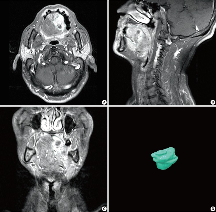
Fig. 1 Magnetic resonance images of a patient with tongue cancer and 3-dimensional reconstructed tongue cancer image. The periphery of tumor is outlined on the magnetic resonance images for tumor volume estimation. (A) Axial image, (B) coronal image, (C) sagittal image, (D) reconstructed image.
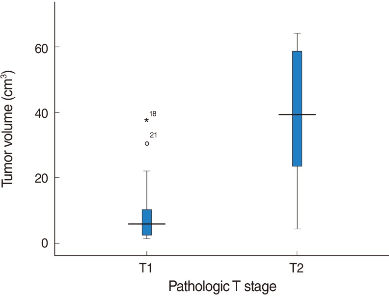
Fig. 2 Relationship between tumor volume and pathologic T stage. A significant relation was found between tumor volume and pathologic T stage. T1=9.49±10.46 cm3, T2=40.06±19.18 cm3, P<0.001.
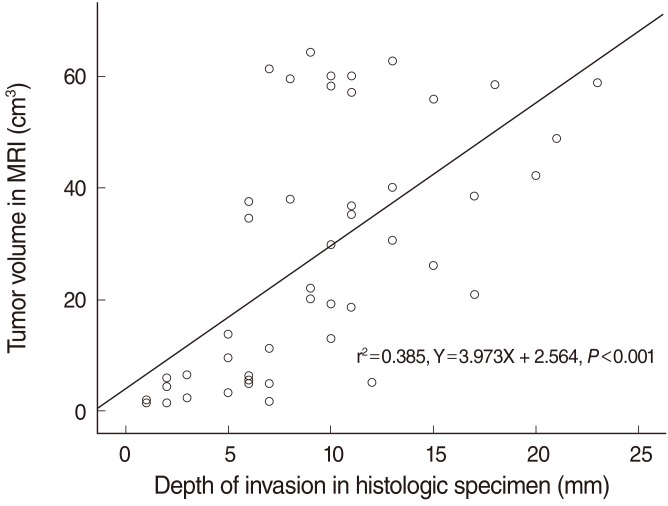
Fig. 3 Relationship between tumor volume and depth of invasion.
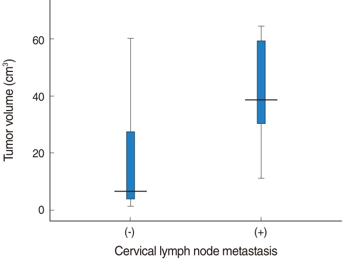
Fig. 4 Relationship between tumor volume and cervical lymph node metastasis. A significant relation was found between tumor volume and the presence of cervical lymph node metastasis (tumor volume in cases with cervical lymph node metastasis=43.61±17.20 cm3, tumor volume in cases without cervical lymph node metastasis=16.91±18.34 cm3; P<0.001).

Fig. 5 Kaplan-Meier 5-year disease specific survival curves according to the tumor volume. There was a statistically significant association between tumor volume and the 5-year disease-specific survival (the 5-year disease-specific survival with a large tumor.
Reference
-
1. Guttman D, Stern Y, Shpitzer T, Ulanovski D, Druzd T, Feinmesser R. Expression of MMP-9, TIMP-1, CD-34 and factor-8 as prognostic markers for squamous cell carcinoma of the tongue. Oral Oncol. 2004; 9. 40(8):798–803. PMID: 15288834.
Article2. Pameijer FA, Balm A, Hilgers F, Muller SH. Variability of tumor volumes in T3-staged head and neck tumors. Head Neck. 1997; 1. 19(1):6–13. PMID: 9030938.
Article3. Johnson CR, Khandelwal SR, Schmidt-Ullrich RK, Ravalese J III, Wazer DE. The influence of quantitative tumour volume measurements on local control in advanced head and neck cancer using concomitant boost accelerated superfractionated irradiation. Int J Radiat Oncol Biol Phys. 1995; 6. 32(3):635–641. PMID: 7790249.4. Brenner DE, Whitley NO, Houk TL, Aisner J, Wiernik P, Whitley J. Volume determinations in computed tomography. JAMA. 1982; 3. 247(9):1299–1302. PMID: 7062546.
Article5. Breiman RS, Beck JW, Korobkin M, Glenny R, Akwari OE, Heaston DK, et al. Volume determinations using computed tomography. AJR Am J Roentgenol. 1982; 2. 138(2):329–333. PMID: 6976739.
Article6. Lee RW, Mancuso AA, Saleh EM, Mendenhall WM, Parsons JT, Million RR. Can pretreatment computed tomography findings predict local control in T3 squamous cell carcinoma of the glottic larynx treated with radiotherapy alone? Int J Radiat Oncol Biol Phys. 1993; 3. 25(4):683–687. PMID: 8454487.
Article7. Freeman DE, Mancuso AA, Parsons JT, Mendenhall WM, Million RR. Irradiation alone for supraglottic larynx carcinoma: can CT findings predict treatment results? Int J Radiat Oncol Biol Phys. 1990; 8. 19(2):485–490. PMID: 2394626.
Article8. Gilbert RW, Birt D, Shulman H, Freeman J, Jenkin D, MacKenzie R, et al. Correlation of tumour volume with local control in laryngeal carcinoma treated by radiation therapy. Ann Otol Rhinol Laryngol. 1987; Sep-Oct. 96(5):514–518. PMID: 3674647.9. Mukherji SK, Mancuso AA, Mendenhall W, Kotzur IM, Kubilis P. Can pretreatment CT predict local control of T2 glottic carcinomas treated with radiation therapy alone? AJNR Am J Neuroradiol. 1995; 4. 16(4):655–662. PMID: 7611018.10. Spiro RH, Huvos AG, Wong GY, Spiro JD, Gnecco CA, Strong EW. Predictive value of tumor thickness in squamous carcinoma confined to the tongue and floor of the mouth. Am J Surg. 1986; 10. 152(4):345–350. PMID: 3766861.
Article11. Kim SH, Cho NH, Kim K, Lee JS, Koo BS, Kim JH, et al. Correlations of oral tongue cancer invasion with matrix metalloproteinases (MMPs) and vascular endothelial growth factor (VEGF) expression. J Surg Oncol. 2006; 3. 93(4):330–337. PMID: 16496371.
Article12. Iwai H, Kyomoto R, Ha-Kawa SK, Lee S, Yamashita T. Magnetic resonance determination of tumour thickness as predictive factor of cervical metastasis in oral tongue carcinoma. Laryngoscope. 2002; 3. 112(3):457–461. PMID: 12148854.13. Brenner DJ. Dose, volume and tumor control predictions in radiotherapy. Int J Radiat Oncol Biol Phys. 1993; 4. 26(1):171–179. PMID: 8482624.14. Johnson CR, Thames HD, Huang DT, Schmidt-Ullrich RK. The tumor volume and clonogen number relationship: Tumor control predictions based upon tumor volume estimates derived from computed tomography. Int J Radiat Oncol Biol Phys. 1995; 9. 33(2):281–287. PMID: 7673015.15. Chua DT, Sham JS, Kwong DL, Tai KS, Wu PM, Lo M, et al. Volumetric analysis of tumor extent in nasopharyngeal carcinoma and correlation with treatment outcome. Int J Radiat Oncol Biol Phys. 1997; 10. 39(3):711–719. PMID: 9336154.
Article16. Willner J, Baier K, Pfreundner L, Flentje M. Tumor volume and local control in primary radiotherapy of nasopharyngeal carcinoma. Acta Oncol. 1999; 38(8):1025–1030. PMID: 10665757.
Article17. Johnson CR, Khandelwal SR, Schmidt-Ullrich RK, Ravalese J 3rd, Wazer DE. The influence of quantitative tumor volume measurements on local control in advanced head and neck cancer using concomitant boost accelerated superfractionated irradiation. Int J Radiat Oncol Biol Phys. 1995; 6. 32(3):635–641. PMID: 7790249.
Article18. Gilbert RW, Birt D, Shulman H, Freeman J, Jenkin D, MacKenzie R, et al. Correlation of tumor volume with local control in laryngeal carcinoma. Ann Otol Rhinol Laryngol. 1987; Sep-Oct. 96(5):514–518. PMID: 3674647.19. Kuriakose MA, Loree TR, Hicks WL, Welch JJ, Wang H, DeLacure MD. Tumour volume estimated by computed tomography as a predictive factor in carcinoma of the tongue. Br J Oral Maxillofac Surg. 2000; 10. 38(5):460–465. PMID: 11010774.
Article20. Chew MH, Khoo JB, Chong VF, Tai BC, Soo KC, Lim DT. Significance of tumour volume measurements in tongue cancer: a novel role in staging. ANZ J Surg. 2007; 8. 77(8):632–637. PMID: 17635274.
Article21. Been MJ, Watkins J, Manz RM, Gentry LR, Leverson GE, Harari PM, et al. Tu mor volume as a prognostic factor in oropharyngeal squamous cell carcinoma treated with primary radiotherapy. Laryngoscope. 2008; 8. 118(8):1377–1382. PMID: 18418275.
- Full Text Links
-
- Actions
-
Cited
- CITED
-
- Close
- Share
-
- Similar articles
-
- Analysis of Influencing Factors on Cervical Lymph Node Metastasis in Early Oral Tongue Cancer
- The Results of Treatment in Clinical N0 Oral Tongue Cancer
- A study of the correlations between the tongue and oral cavity volume in the skeletal mandibular prognathism
- Clinical Efficacy of Primary Tumor Volume Measurements: Comparison of Different Primary Sites
- A case of tongue volume measurement with the plaster tongue model for partial glossectomy in macroglossia
