Korean J Pain.
2015 Apr;28(2):105-108. 10.3344/kjp.2015.28.2.105.
Detection Rate of Intravascular Injections during Cervical Medial Branch Blocks: A Comparison of Digital Subtraction Angiography and Static Images from Conventional Fluoroscopy
- Affiliations
-
- 1Department of Anesthesiology and Pain Medicine, School of Dentistry, Kyungpook National University, Daegu, Korea.
- 2School of Medicine, Keimyung University, Daegu, Korea. mandell@naver.com
- KMID: 2278268
- DOI: http://doi.org/10.3344/kjp.2015.28.2.105
Abstract
- BACKGROUND
The most definitive diagnosis of neck pain caused by facet joints can be obtained through cervical medial branch blocks (CMBBs). However, intravascular injections need to be carefully monitored, as they can increase the risk of false-negative blocks when diagnosing cervical facet joint syndrome. In addition, intravascular injections can cause neurologic deficits such as spinal infarction or cerebral infarction. Digital subtraction angiography (DSA) is a radiological technique that can be used to clearly visualize the blood vessels from surrounding bones or dense soft tissues. The purpose of this study was to compare the rate of detection of intravascular injections during CMBBs using DSA and static images obtained through conventional fluoroscopy.
METHODS
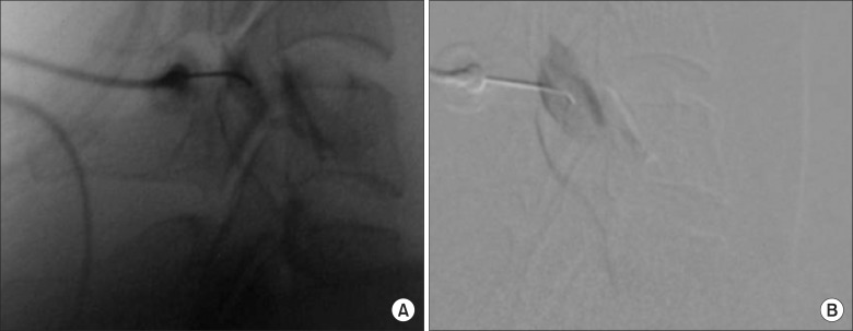
Seventy-two patients were included, and a total of 178 CMBBs were performed. The respective incidences of intravascular injections during CMBBs using DSA and static images from conventional fluoroscopy were measured.
RESULTS
A total of 178 CMBBs were performed on 72 patients. All cases of intravascular injections evidenced by the static images were detected by the DSAs. The detection rate of intravascular injections was higher from DSA images than from static images (10.7% vs. 1.7%, P < 0.001).
CONCLUSIONS
According to these findings, the use of DSA can improve the detection rate of intravascular injections during CMBBs. The use of DSA may therefore lead to an increase in the diagnostic and therapeutic value of CMBBs. In addition, it can decrease the incidence of potential side effects during CMBBs.
Keyword
MeSH Terms
Figure
Cited by 1 articles
-
Effect of needle type on intravascular injection in transforaminal epidural injection: a meta-analysis
Jae Yun Kim, Soo Nyoung Kim, Chulmin Park, Ho Young Lim, Jae Hun Kim
Korean J Pain. 2019;32(1):39-46. doi: 10.3344/kjp.2019.32.1.39.
Reference
-
1. Aprill C, Bogduk N. The prevalence of cervical zygapophyseal joint pain. A first approximation. Spine (Phila Pa 1976). 1992; 17:744–747. PMID: 1502636.
Article2. Falco FJ, Erhart S, Wargo BW, Bryce DA, Atluri S, Datta S, et al. Systematic review of diagnostic utility and therapeutic effectiveness of cervical facet joint interventions. Pain Physician. 2009; 12:323–344. PMID: 19305483.3. Manchikanti L, Boswell MV, Singh V, Benyamin RM, Fellows B, Abdi S, et al. Comprehensive evidence-based guidelines for interventional techniques in the management of chronic spinal pain. Pain Physician. 2009; 12:699–802. PMID: 19644537.4. Rathmell JP, Lake T, Ramundo MB. Infectious risks of chronic pain treatments: injection therapy, surgical implants, and intradiscal techniques. Reg Anesth Pain Med. 2006; 31:346–352. PMID: 16857554.
Article5. Verrills P, Mitchell B, Vivian D, Nowesenitz G, Lovell B, Sinclair C. The incidence of intravascular penetration in medial branch blocks: cervical, thoracic, and lumbar spines. Spine (Phila Pa 1976). 2008; 33:E174–E177. PMID: 18344846.6. Boswell MV, Trescot AM, Datta S, Schultz DM, Hansen HC, Abdi S, et al. Interventional techniques: evidence-based practice guidelines in the management of chronic spinal pain. Pain Physician. 2007; 10:7–111. PMID: 17256025.7. Brouwers PJ, Kottink EJ, Simon MA, Prevo RL. A cervical anterior spinal artery syndrome after diagnostic blockade of the right C6-nerve root. Pain. 2001; 91:397–399. PMID: 11275398.
Article8. Heckmann JG, Maihöfner C, Lanz S, Rauch C, Neundörfer B. Transient tetraplegia after cervical facet joint injection for chronic neck pain administered without imaging guidance. Clin Neurol Neurosurg. 2006; 108:709–711. PMID: 16102894.
Article9. Karasek M, Bogduk N. Temporary neurologic deficit after cervical transforaminal injection of local anesthetic. Pain Med. 2004; 5:202–205. PMID: 15209975.
Article10. Tiso RL, Cutler T, Catania JA, Whalen K. Adverse central nervous system sequelae after selective transforaminal block: the role of corticosteroids. Spine J. 2004; 4:468–474. PMID: 15246308.
Article11. Smuck M, Fuller BJ, Chiodo A, Benny B, Singaracharlu B, Tong H, et al. Accuracy of intermittent fluoroscopy to detect intravascular injection during transforaminal epidural injections. Spine (Phila Pa 1976). 2008; 33:E205–E210. PMID: 18379390.
Article12. McLean JP, Sigler JD, Plastaras CT, Garvan CW, Rittenberg JD. The rate of detection of intravascular injection in cervical transforaminal epidural steroid injections with and without digital subtraction angiography. PM R. 2009; 1:636–642. PMID: 19627957.
Article13. Lee MH, Yang KS, Kim YH, Jung HD, Lim SJ, Moon DE. Accuracy of live fluoroscopy to detect intravascular injection during lumbar transforaminal epidural injections. Korean J Pain. 2010; 23:18–23. PMID: 20552068.
Article14. Jasper JF. Role of digital subtraction fluoroscopic imaging in detecting intravascular injections. Pain Physician. 2003; 6:369–372. PMID: 16880884.15. Sehgal N, Dunbar EE, Shah RV, Colson J. Systematic review of diagnostic utility of facet (zygapophysial) joint injections in chronic spinal pain: an update. Pain Physician. 2007; 10:213–228. PMID: 17256031.
- Full Text Links
- Actions
-
Cited
- CITED
-
- Close
- Share
- Similar articles
-
- Digital subtraction angiography vs. real-time fluoroscopy for detection of intravascular injection during transforaminal epidural block
- Accuracy of Live Fluoroscopy to Detect Intravascular Injection During Lumbar Transforaminal Epidural Injections
- Comparison of incidence of intravascular injections during transforaminal epidural steroid injection using different needle types
- Radiologic consideration of intra-arterial digital subtraction angiography
- Intravascular injection in cervical medial branch block: an evaluation of 361 injections


