Ultrasound-guided Aspiration of the Iatrogenic Pneumothorax Caused by Paravertebral Block: A Case Report
- Affiliations
-
- 1Department of Anesthesiology and Pain Medicine, College of Medicine, The Catholic University of Korea, Seoul, Korea. demoon@catholic.ac.kr
- KMID: 2278113
- DOI: http://doi.org/10.3344/kjp.2012.25.1.33
Abstract
- Thoracic paravertebral block is performed for the treatment of patients with chronic pain, such as complex regional pain syndrome (CRPS) and post-herpetic neuralgia. Thoracic paravertebral block can result in iatrogenic pneumothorax. Because pneumothorax can develop into medical emergencies and needle aspiration or chest tube placement may be needed, early diagnosis is very important. Recently, thoracic ultrasonography has begun to be used to diagnose pneumothorax. In addition, ultrasound-guided aspiration can be an accurate and safe technique for treatment of pneumothorax, as the needle position can be followed in real time. We report a case of iatrogenic pneumothorax following thoracic paravertebral block for the treatment of chronic pain due to CRPS, treated successfully by ultrasound-guided aspiration.
Keyword
Figure
-
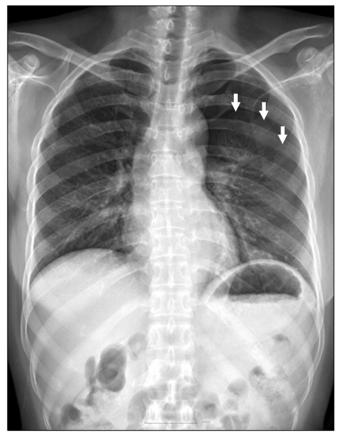
Fig. 1 Initial chest x-ray shows pneumothorax in left hemithorax (white arrow) and partially collapsed left lung.
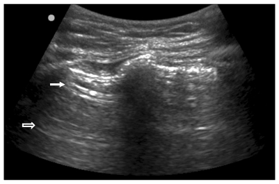
Fig. 2 Initial ultrasound images of the second intercostal space. White arrow indicates parietal pleura without lung sliding sign. Blanked arrow indicates horizontal artifacts.
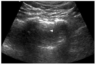
Fig. 3 Ultrasound image during aspiration of air in the interpleural space. Arrowhead indicates the needle tip bellow the parietal pleura.
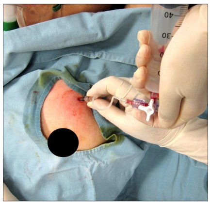
Fig. 4 A picture which shows aspiration using 18 gauge angiocatheter, 3 ways valve and 50 ml syringe. A angiocatheter is placed in the second intercostal area. A large block dot indicate the nipple of patient.

Fig. 5 Chest-x ray after air aspiration shows shows decreased size of pneumothorax (arrowhead).

Fig. 6 Ultrasound image after air aspiration. This shows that lung sliding sign is seen of the plural line (white arrow) and comet-tail artifact from the pleural line to the edge of the screen (arrowhead).
Cited by 1 articles
-
Ultrasound Sonography at the Pain Clinic in Korea: Past, Present and Future
Ji Yong Park
Korean J Pain. 2013;26(1):1-2. doi: 10.3344/kjp.2013.26.1.1.
Reference
-
1. Lönnqvist PA, MacKenzie J, Soni AK, Conacher ID. Paravertebral blockade. Failure rate and complications. Anaesthesia. 1995; 50:813–815. PMID: 7573876.2. Alrajhi K, Woo MY, Vaillancourt C. Test characteristics of ultrasonography for the detection of pneumothorax: a systematic review and meta-analysis. Chest [serial on the Internet]. 2011. 8. [2011 Aug 25]. Available at http://chestjournal.chestpubs.org/content/early/2011/08/24/chest.11-0131.3. Lichtenstein DA, Mezière G, Lascols N, Biderman P, Courret JP, Gepner A, et al. Ultrasound diagnosis of occult pneumothorax. Crit Care Med. 2005; 33:1231–1238. PMID: 15942336.
Article4. Novak-Jankovic V. Update on thoracic paravertebral blocks. Coll Antropol. 2011; 35:595–598. PMID: 21755736.5. Medford AR, Entwisle JJ. Indications for thoracic ultrasound in chest medicine: an observational study. Postgrad Med J. 2010; 86:8–11. PMID: 20065335.
Article6. Lichtenstein D, Mezière G, Biderman P, Gepner A. The comet-tail artifact: an ultrasound sign ruling out pneumothorax. Intensive Care Med. 1999; 25:383–388. PMID: 10342512.
Article7. Lichtenstein DA, Menu Y. A bedside ultrasound sign ruling out pneumothorax in the critically ill. Lung sliding. Chest. 1995; 108:1345–1348. PMID: 7587439.
Article8. Rowan KR, Kirkpatrick AW, Liu D, Forkheim KE, Mayo JR, Nicolaou S. Traumatic pneumothorax detection with thoracic US: correlation with chest radiography and CT--initial experience. Radiology. 2002; 225:210–214. PMID: 12355007.
Article9. Yang SY, Sung CH, Yoon KJ, Kim SH, Moon SW, Moon DE. Iatrogenic pneumothorax after thoracic paravertebral block: a report of 2 cases. J Korean Pain Soc. 2003; 16:273–277.
- Full Text Links
-
- Actions
-
Cited
- CITED
-
- Close
- Share
-
- Similar articles
-
- Iatrogenic Pneumothorax after Thoracic Paravertebral Block: A report of 2 cases
- Treatment of Isolated Sternal Fracture with Ultrasound-Guided Paravertebral Nerve Block: a Case Report and Literature Review
- Ultrasound Guided Thoracic Paravertebral Space Block for Chronic Intractable Upper Back Pain
- Pneumothorax
- Pneumothorax after Interventional Muscle and Soft Tissue Stimulation Therapy : A case report
