Retroperitoneal recurrence of uterine smooth muscle tumor of uncertain malignant potential as leiomyosarcoma
- Affiliations
-
- 1Department of Obstetrics and Gynecology, Inha University College of Medicine, Incheon, Korea. mdpjw1216@gmail.com
- 2Department of Pathology, Inha University College of Medicine, Incheon, Korea.
- KMID: 2274213
- DOI: http://doi.org/10.5468/KJOG.2012.55.12.996
Abstract
- A 57-year-old Korean woman visited 37 months after initial surgery with fetal head sized pelvic mass. Initially, she underwent total abdominal hysterectomy with bilateral salpingo-oophorectomy, omentectomy, and para-aortic lymph node biopsy. The initial pathological report showed uterine smooth muscle tumors of uncertain malignant potential (STUMP). She had not received adjuvant therapy. Transvaginal ultrasound revealed multiple solid masses in the pelvic cavity. Abdomino-pelvic computed tomography scan revealed heterogeneous lobulated rounding masses. Tumor markers were all within the normal range. She underwent explorative laparotomy. The pathological diagnosis was epitheloid leiomyosarcoma. Patients with STUMP should be counseled regarding the potential for recurrence as leiomyosarcoma, and may require closer surveillance than a yearly.
MeSH Terms
Figure
-
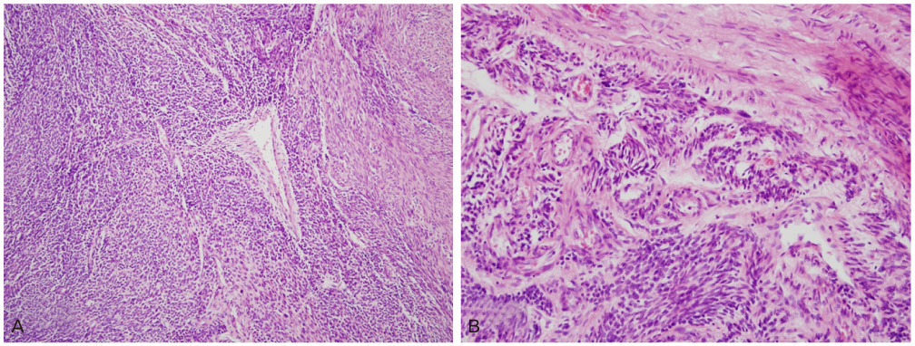
Fig. 1 (A) The tumor was characterized by smooth muscle tumor with high celluarity not accompanied by tumor cell necrosis or significant nuclear atypia (H&E, ×100). (B) However, a minor focal area exhibited moderate to severe nuclear atypia with high mitotic figures (H&E, ×200).
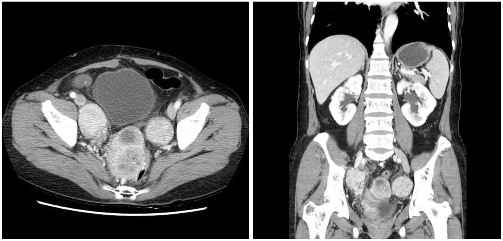
Fig. 2 Abdominopelvic computed tomography shows 4 × 4, 4 × 6, 6 × 7 cm sized heterogeneous lobulated rounding masses, suggestive recurrence of pelvic cavity, and lymphatic dissemination of rounding masses along iliac vessels.
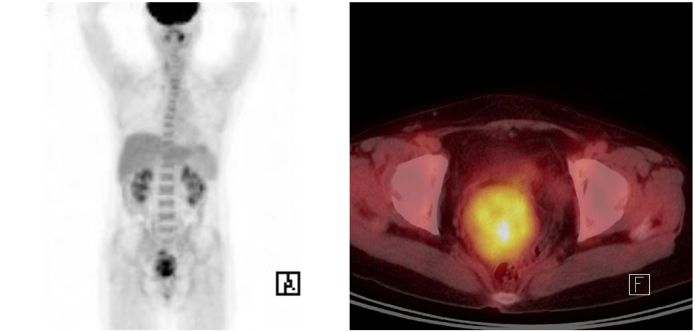
Fig. 3 Positron emission tomography/computed tomography showed several recurrent masses with abnormal fluorodeoxyglucose uptake in the pelvic cavity.
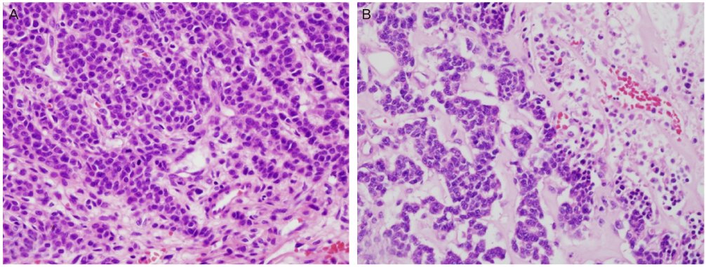
Fig. 4 (A) The tumor consists of poorly differentiated cells with round to oval hyperchromatic nuclei and frequent mitotic figures (H&E, ×400). (B) In some areas showed geographic necrosis (H&E, ×400).
Reference
-
1. Guntupalli SR, Ramirez PT, Anderson ML, Milam MR, Bodurka DC, Malpica A. Uterine smooth muscle tumor of uncertain malignant potential: a retrospective analysis. Gynecol Oncol. 2009. 113:324–326.2. Berretta R, Rolla M, Merisio C, Giordano G, Nardelli GB. Uterine smooth muscle tumor of uncertain malignant potential: a three-case report. Int J Gynecol Cancer. 2008. 18:1121–1126.3. Shapiro A, Ferenczy A, Turcotte R, Bruchim I, Gotlieb WH. Uterine smooth-muscle tumor of uncertain malignant potential metastasizing to the humerus as a high-grade leiomyosarcoma. Gynecol Oncol. 2004. 94:818–820.4. D'Angelo E, Prat J. Uterine sarcomas: a review. Gynecol Oncol. 2010. 116:131–139.5. Bell SW, Kempson RL, Hendrickson MR. Problematic uterine smooth muscle neoplasms. A clinicopathologic study of 213 cases. Am J Surg Pathol. 1994. 18:535–558.6. Hart WR. Problematic uterine smooth muscle neoplasms. Am J Surg Pathol. 1997. 21:252–255.7. Amant F, Moerman P, Vergote I. Report of an unusual problematic uterine smooth muscle neoplasm, emphasizing the prognostic importance of coagulative tumor cell necrosis. Int J Gynecol Cancer. 2005. 15:1210–1212.8. Ip PP, Cheung AN, Clement PB. Uterine smooth muscle tumors of uncertain malignant potential (STUMP): a clinicopathologic analysis of 16 cases. Am J Surg Pathol. 2009. 33:992–1005.9. Atkins KA, Arronte N, Darus CJ, Rice LW. The Use of p16 in enhancing the histologic classification of uterine smooth muscle tumors. Am J Surg Pathol. 2008. 32:98–102.10. Slik K, El Rishi F, Bulugma M. Uterine smooth muscle tumors: clinicopathological evaluation. Free communication oral presentations. Int J Gynecol Obstet. 2009. 107(S2):S393–S396.
- Full Text Links
-
- Actions
-
Cited
- CITED
-
- Close
- Share
-
- Similar articles
-
- A Case of Cutaneous Smooth Muscle Tumor of Uncertain Malignant Potential
- A case of retroperitoneal huge smooth muscle tumor misleading to ovarian cancer
- A Clinical - Pathological Study of Uterine Smooth Muscle Tumor of Uncertain Malignant Potential
- Transvaginal Color Doppler Sonographv of Uterine Smooth Muscle Tumors: Prediction of the Extent of Degenerative Change and Differentiation of Leiomyosarcoma from Myoma
- Uterine smooth muscle tumor of uncertain malignant potential: fertility and clinical outcomes
