Malignant mixed Mullerian tumor of uterine cervix: A case of very poor prognosis
- Affiliations
-
- 1Department of Obstetrics and Gynecology, CHA Gangnam Medical Center, CHA University, Seoul, Korea. sjseongcheil@yahoo.co.kr
- 2Department of Pathology, CHA Gangnam Medical Center, CHA University, Seoul, Korea.
- KMID: 2274212
- DOI: http://doi.org/10.5468/KJOG.2012.55.12.990
Abstract
- Malignant mixed Mullerian tumor (MMMT) is a type of cancer that contains two cell types known as the adenocarcinoma and sarcoma. MMMT is a rare malignant cancer of the genital tract and particularly the prevalence of MMMT in the cervix is extremely rare. Due to the rare incidence of MMMT in cervix, its treatment has unestablished guidelines and has unclear prognosis. Based on the previous data, the prognosis of MMMT in the cervix is better than the prognosis of uterine MMMT. Here, we are reporting a case of cervix MMMT that is rapidly progressive with an extremely poor prognosis, which contradicts the previous data.
Figure
-
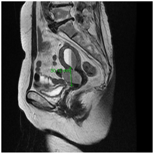
Fig. 1 Magnetic resonance imaging finding. 2.6 × 3.4 × 3.0 cm sized enhancing cervical mass.
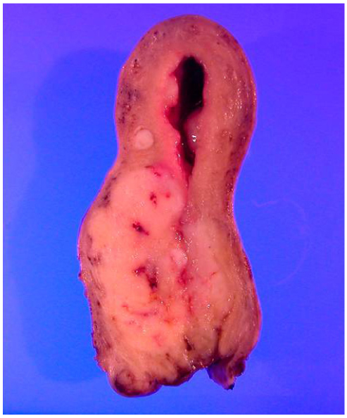
Fig. 2 In the uterine cervix and the lower segment of the uterus, a beige colored, 5.4 × 3 cm sized solid mass with ill-defined boundaries is observed.
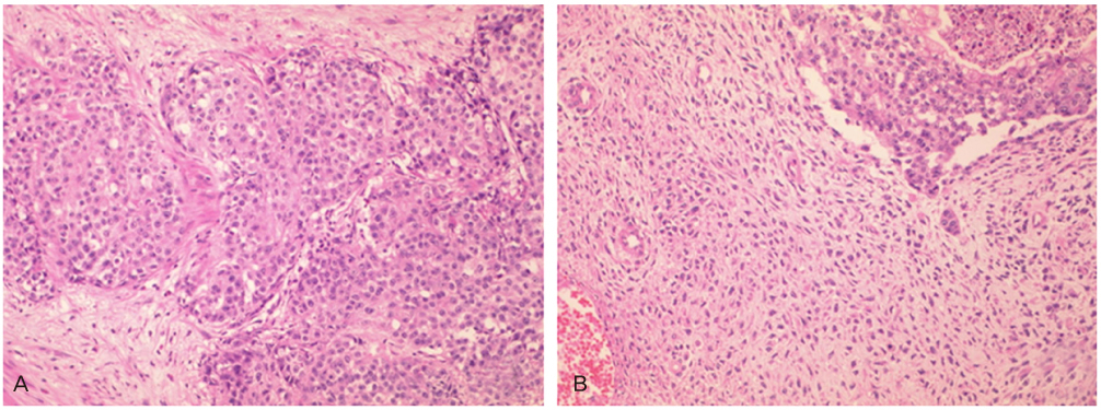
Fig. 3 The epithelial cell component is mostly composed of undifferentiated carcinoma cells (A, H&E, ×200) and the stromal cell component is mainly composed of undifferentiated sarcoma cells (B, H&E, ×200). Pleomorphism and mitosis is observed in both of the components which is consistent with malignant mixed Müllerian tumor.

Fig. 4 Adenosquamous cell carcinoma, undifferentiated sarcoma and chondylosarcoma are partly observed (A, H&E, ×200). After immunohistochemical stain adenosquamous cell carcinoma was cytokeratin positive (B, cytokeratin, ×200) and undifferentiated sarcoma and chondylosarcoma was vimentin positive (C, vimentin, ×200).
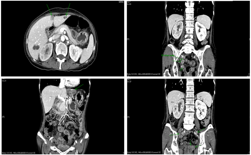
Fig. 5 Abdominopelvic computed tomography images 1 month after operation. Metastatic masses were found at liver, pelvic cavity, and multiple lymph nodes. Also, right hydronephrosis and hydroureter were checked.

Fig. 6 Preoperative and postoperative positron emission tomography-computed tomography (PET-CT) findings. (A) Preoperative PET-CT images. (B) Postoperative 1 month later PET-CT images.
Reference
-
1. Brown J, Broaddus R, Koeller M, Burke TW, Gershenson DM, Bodurka DC. Sarcomatoid carcinoma of the cervix. Gynecol Oncol. 2003. 90:23–28.2. Rodrigues L, Santana I, Cunha T, Felix A, Freire J, Cabral I. Sarcomatoid squamous cell carcinoma of the uterine cervix: case report. Eur J Gynaecol Oncol. 2000. 21:287–289.3. Sharma NK, Sorosky JI, Bender D, Fletcher MS, Sood AK. Malignant mixed mullerian tumor (MMMT) of the cervix. Gynecol Oncol. 2005. 97:442–445.4. Clement PB, Zubovits JT, Young RH, Scully RE. Malignant mullerian mixed tumors of the uterine cervix: a report of nine cases of a neoplasm with morphology often different from its counterpart in the corpus. Int J Gynecol Pathol. 1998. 17:211–222.5. Manolitsas TP, Wain GV, Williams KE, Freidlander M, Hacker NF. Multimodality therapy for patients with clinical Stage I and II malignant mixed Mullerian tumors of the uterus. Cancer. 2001. 91:1437–1443.6. Piver MS, Lurain JR. Coppleson M, editor. Uterine sarcomas: clinical features and management. Gynecologic oncology: fundamental principles and clinical practice. 1981. Vol. 2. Edinburgh: Churchill Livingstone;608–618.7. Bansal S, Lewin SN, Burke WM, Deutsch I, Sun X, Herzog TJ, et al. Sarcoma of the cervix: natural history and outcomes. Gynecol Oncol. 2010. 118:134–138.8. Sunwoo J, Cho IS, Jeon S, Bae DH, Shin YW, Kim CJ, et al. A case of malignant mixed mullerian tumor (MMMT) of the uterine cervix. Korean J Obstet Gynecol. 2008. 51:350–354.
- Full Text Links
-
- Actions
-
Cited
- CITED
-
- Close
- Share
-
- Similar articles
-
- A case of malignant Mixed Mullerian Tumor (MMMT) of the uterine cervix
- Malignant Mixed Mullerian Tumor Arising from the Uterine Cervix: A Case Report
- Malignant Mixed Mullerian Tumor of the Fallopian Tube: Report of a Case
- Two Case of Malignant Mixed Mullerian Tumors
- A Case of Malignant Mixed Mullerian Tumor of the Fallopian Tube
