Korean J Obstet Gynecol.
2012 Nov;55(11):853-858. 10.5468/KJOG.2012.55.11.853.
A case of primary fallopian tube carcinoma diagnosed radiologically before operation
- Affiliations
-
- 1Department of Obstetrics and Gynecology, Keimyung University School of Medicine, Daegu, Korea. ksh1999@dsmc.or.kr
- 2Department of Pathology, Keimyung University School of Medicine, Daegu, Korea.
- KMID: 2274156
- DOI: http://doi.org/10.5468/KJOG.2012.55.11.853
Abstract
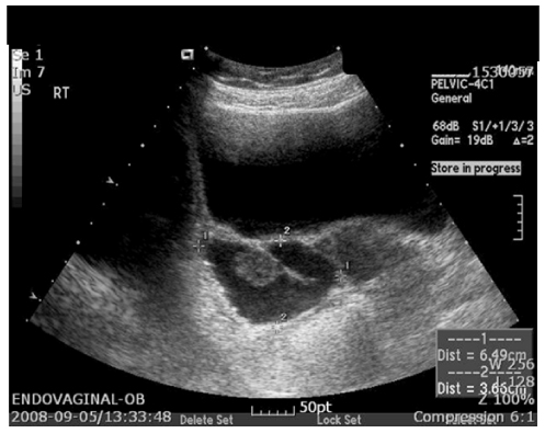
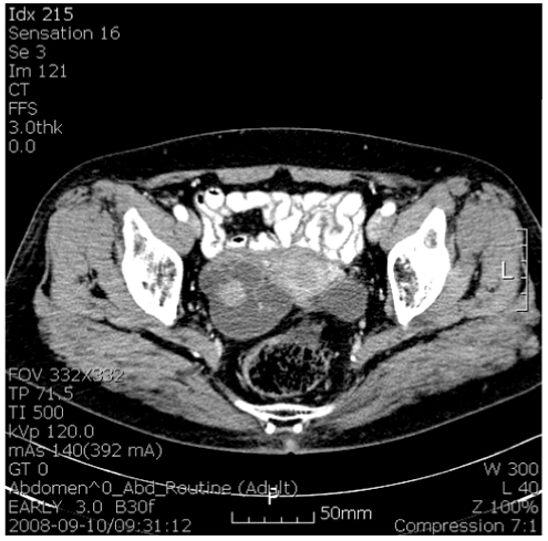
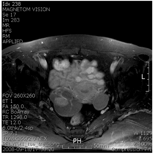
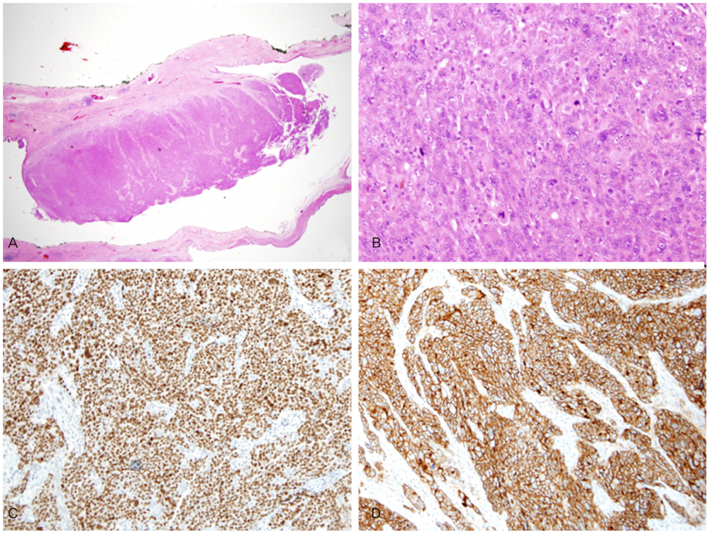
- Primary fallopian tube carcinoma is one of the rarest gynecological malignancies, accounting for 0.18% to 1.6% of all malignant neoplasms of the female reproductive tract. Preoperative diagnosis was difficult due to nonspecific symptoms and signs. This case of primary tubal cancer was diagnosed preoperatively on the basis of ultrasonography, computed tomography and magnetic resonance imaging. We have experienced a case of primary fallopian tube carcinoma before operation and so report with brief review of the literature.
Figure
Reference
-
1. Rosenblatt KA, Weiss NS, Schwartz SM. Incidence of malignant fallopian tube tumors. Gynecol Oncol. 1989. 35:236–239.2. Dodson MG, Ford JH Jr, Averette HE. Clinical aspects of fallopian tube carcinoma. Obstet Gynecol. 1970. 36:935–939.3. Sedlis A. Primary carcinoma of the fallopian tube. Obstet Gynecol Surv. 1961. 16:209–226.4. Johnston GA Jr. Primary malignancy of the fallopian tube: a clinical review of 13 cases. J Surg Oncol. 1983. 24:304–309.5. Lootsma-Miklosova E, Aalders JG, Willemse PH, de Bruijn HW. Levels of CA 125 in patients with recurrent carcinoma of the fallopian tube: two case histories. Eur J Obstet Gynecol Reprod Biol. 1987. 24:231–235.6. Kurjak A, Kupesic S, Ilijas M, Sparac V, Kosuta D. Preoperative diagnosis of primary fallopian tube carcinoma. Gynecol Oncol. 1998. 68:29–34.7. Kawakami S, Togashi K, Kimura I, Nakano Y, Koshiyama M, Takakura K, et al. Primary malignant tumor of the fallopian tube: appearance at CT and MR imaging. Radiology. 1993. 186:503–508.8. Kahng YR, Kim JK, Cho KS. Primary malignant tumor of the fallopian tube: CT and MR features. J Korean Radiol Soc. 2001. 45:393–397.9. Subramanyam BR, Raghavendra BN, Whalen CA, Yee J. Ultrasonic features of fallopian tube carcinoma. J Ultrasound Med. 1984. 3:391–393.10. Roberts JA, Lifshitz S. Primary adenocarcinoma of the fallopian tube. Gynecol Oncol. 1982. 13:301–308.11. Hu CY, Taymor ML, Hertig AT. Primary carcinoma of the fallopian tube. Am J Obstet Gynecol. 1950. 59:58–67.12. Kim TS, Jang HS, Han DG, Kim SL, Choi YC. A case of primary adenosquamous carcinoma of the fallopian tube. Korean J Obstet Gynecol. 1994. 37:1865–1871.13. Oh YS, Yi SW, Huh CY, Kim SB. Two cases of primary carcinoma of the fallopian tube. Korean J Obstet Gynecol. 1999. 42:1849–1853.14. Benedet JL, Bender H, Jones H 3rd, Ngan HY, Pecorelli S. FIGO Committee on Gynecologic Oncology. FIGO staging classifications and clinical practice guidelines in the management of gynecologic cancers. Int J Gynaecol Obstet. 2000. 70:209–262.15. Jung YJ, Chi KS, Kim JS, Kim KW, Kim DG, Yang HS, et al. 3 cases of primary tubal cancer incidentally diagnosed after benign gynecologic operation. Korean J Obstet Gynecol. 2006. 49:1779–1787.


