A case of arthrogryposis multiplex congenita associated with maternal septate uterus
- Affiliations
-
- 1Department of Obstetrics and Gynecology, Catholic University of Daegu School of Medicine, Daegu, Korea. magu815@cu.ac.kr
- 2Department of Pathology, Catholic University of Daegu School of Medicine, Daegu, Korea.
- KMID: 2274141
- DOI: http://doi.org/10.5468/KJOG.2012.55.9.669
Abstract
- Arthrogryposis multiplex congenita (AMC) is a congenital disorder showing multiple joint contractures. Although there are characteristic features in morphology, it is difficult to diagnose prenatally by ultrasonography. The causes of AMC include abnormalities of central nervous system, myopathies, connective tissue disorders, and intrauterine environmental factors such as oligohydramnios or uterine anomalies. We describe a case of prenatally diagnosed and postnatally evaluated AMC associated with maternal septate uterus.
MeSH Terms
Figure
-
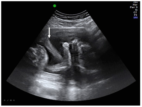
Fig. 1 Coronal scan of gravid uterus shows that a thick septum is extended to lower uterine cavity. The head and upper portion of the fetus is located in the right uterine cavity and lower portion in the left cavity (arrow, septum).
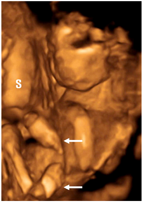
Fig. 2 3-D scan image of multiple joint contractures (S, setum; arrow, wrist deformity and club foot).
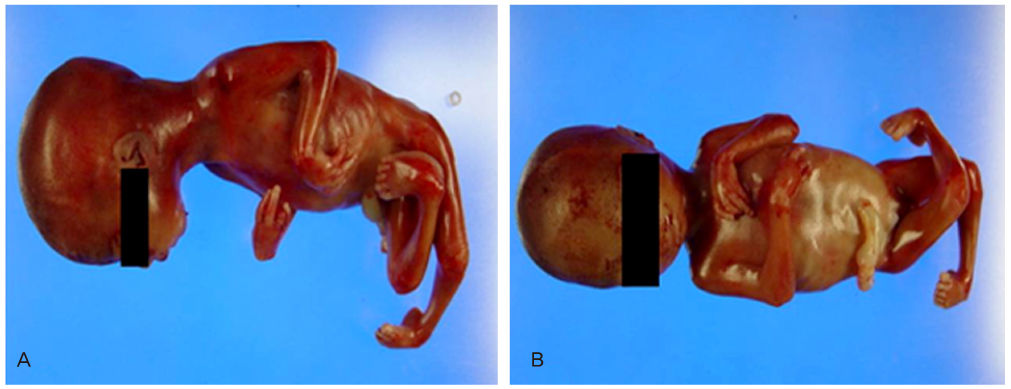
Fig. 3 Gross appearances of the fetus with multiple joint contractures corresponding arhtrogryposis multiplex congenita.
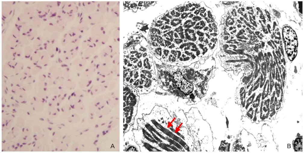
Fig. 4 (A) High power microscopic findings of calf muscle shows mild size variation of skeletal muscle fiber and interstitial edema (H&E, ×400). (B) Electron microscope shows well preserved myofbrils and sarcomeres (arrow). There is no abnormal mitochondria nor cytoplasmic inclusion (×1,500).
Reference
-
1. Hall JG. Genetic aspects of arthrogryposis. Clin Orthop Relat Res. 1985. (194):44–53.2. Fassier A, Wicart P, Dubousset J, Seringe R. Arthrogryposis multiplex congenita. Long-term follow-up from birth until skeletal maturity. J Child Orthop. 2009. 3:383–390.3. Chung IH, Park BM. A case of Arthrogryposis multiplex congenita. J Korean Med Assoc. 1961. 4:62–64.4. Mennen U, van Heest A, Ezaki MB, Tonkin M, Gericke G. Arthrogryposis multiplex congenita. J Hand Surg Br. 2005. 30:468–474.5. Lin IW, Chueh HY, Chang SD, Cheng PJ. The application of three-dimensional ultrasonography in the prenatal diagnosis of arthrogryposis. Taiwan J Obstet Gynecol. 2008. 47:75–78.6. Hall JG. Arthrogryposis multiplex congenita: etiology, genetics, classification, diagnostic approach, and general aspects. J Pediatr Orthop B. 1997. 6:159–166.7. Vuopala K, Leisti J, Herva R. Lethal arthrogryposis in Finland--a clinico-pathological study of 83 cases during thirteen years. Neuropediatrics. 1994. 25:308–315.8. Gordon N. Arthrogryposis multiplex congenita. Brain Dev. 1998. 20:507–511.9. Zori RT, Gardner JL, Zhang J, Mullan MJ, Shah R, Osborn AR, et al. Newly described form of X-linked arthrogryposis maps to the long arm of the human X chromosome. Am J Med Genet. 1998. 78:450–454.10. Bamshad M, Van Heest AE, Pleasure D. Arthrogryposis: a review and update. J Bone Joint Surg Am. 2009. 91:Suppl 4. 40–46.11. Hardwick JC, Irvine GA. Obstetric care in arthrogryposis multiplex congenita. BJOG. 2002. 109:1303–1304.12. Kang PB, Lidov HG, David WS, Torres A, Anthony DC, Jones HR, et al. Diagnostic value of electromyography and muscle biopsy in arthrogryposis multiplex congenita. Ann Neurol. 2003. 54:790–795.
