Korean J Obstet Gynecol.
2011 Jun;54(6):308-313. 10.5468/KJOG.2011.54.6.308.
Ultrasonographic follow-up of involuting placenta in advanced abdominal pregnancy diagnosed by magnetic resonance imaging
- Affiliations
-
- 1Department of Obstetrics and Gynecology, Chungbuk National University Hospital; Medical Research Institute, Chungbuk National University, Cheongju, Korea. jeongmed@chungbuk.ac.kr
- KMID: 2274040
- DOI: http://doi.org/10.5468/KJOG.2011.54.6.308
Abstract
- We successfully treated a case of advanced abdominal pregnancy which was diagnosed with ultrasonography and magnetic resonance imaging in a woman of 20th week of pregnancy who did not receive any prenatal care. It is advisable to leave the placenta in situ because removing the placenta may be associated with severe hemorrhage. Furthermore, identification of uterine wall around the fetus and the placenta is very important in diagnosing abdominal pregnancy.
MeSH Terms
Figure
-
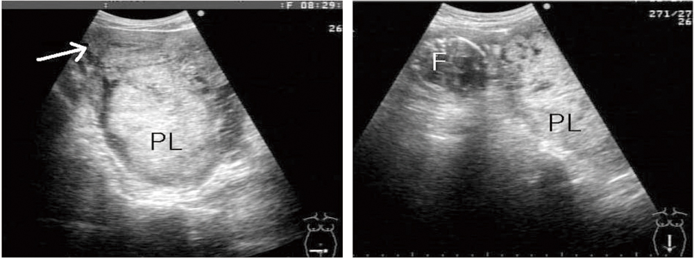
Fig. 1 Transabdominal ultrasonography shows empty uterus (white arrow), extrauterine placenta (PL) and fetal head (F).
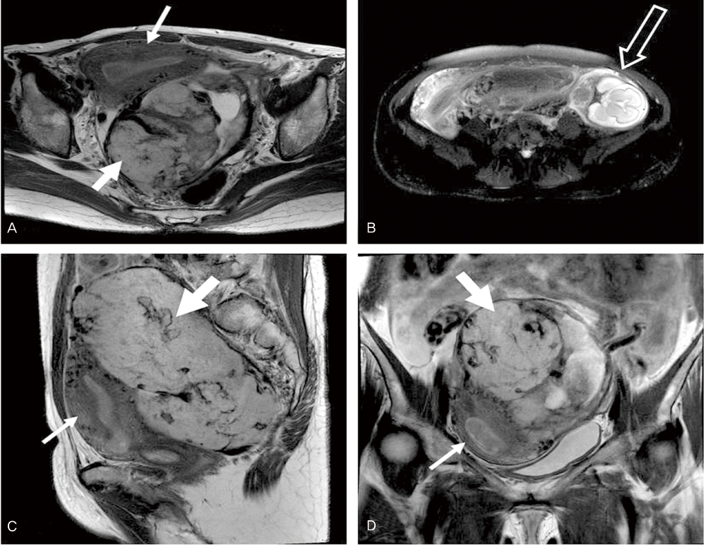
Fig. 2 T2 weighted magnetic resonance imaging shows empty uterus (thin arrow), extrauterine placenta (thick arrow) and the fetus (black arrow). (A) and (B) Axial images. (C) Sagital image. (D) Coronal image.
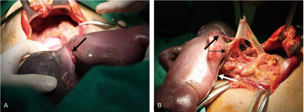
Fig. 3 (A) Omental band strangulates the fetal neck (thick arrow). (B) Omentum attached to the right fetal shoulder (thin arrow) and anterior chest (white arrow).
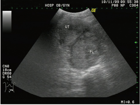
Fig. 4 Involuting placenta (124×83.5×103 mm) still remains in the posterior cul-de sac on 110th postoperative day. The placenta was 160×84×110 mm in size before operation. UT, uterus; PL, placenta.
Reference
-
1. Atrash HK, Friede A, Hogue CJ. Abdominal pregnancy in the United States: frequency and maternal mortality. Obstet Gynecol. 1987. 69:333–337.2. Martin JN Jr, McCaul JF 4th. Emergent management of abdominal pregnancy. Clin Obstet Gynecol. 1990. 33:438–447.3. Kang IS, Jin KS, Hong JG, Heo JY, Kim SB. A case of abdominal pregnancy. Korean J Obstet Gynecol. 1995. 38:895–898.4. Costa SD, Presley J, Bastert G. Advanced abdominal pregnancy. Obstet Gynecol Surv. 1991. 46:515–525.5. Weiss RE, Stone NN. Persistent maternal hydronephrosis after intra-abdominal pregnancy. J Urol. 1994. 152:1196–1198.6. Harris MB, Angtuaco T, Frazier CN, Mattison DR. Diagnosis of a viable abdominal pregnancy by magnetic resonance imaging. Am J Obstet Gynecol. 1988. 159:150–151.7. Rahman MS, Al-Suleiman SA, Rahman J, Al-Sibai MH. Advanced abdominal pregnancy: observations in 10 cases. Obstet Gynecol. 1982. 59:366–372.8. Akhan O, Cekirge S, Senaati S, Besim A. Sonographic diagnosis of an abdominal ectopic pregnancy. AJR Am J Roentgenol. 1990. 155:197–198.9. Alexander MC, Horger EO 3rd. Early diagnosis of abdominal pregnancy by ultrasound. J Clin Ultrasound. 1983. 11:45–48.10. Hage ML, Wall LL, Killam A. Expectant management of abdominal pregnancy. A report of two cases. J Reprod Med. 1988. 33:407–410.11. Martin JN Jr, Sessums JK, Martin RW, Pryor JA, Morrison JC. Abdominal pregnancy: current concepts of management. Obstet Gynecol. 1988. 71:549–557.12. Valenzano M, Nicoletti L, Odicino F, Cocuccio S, Lorenzi P, Ragni N. Five-year follow-up of placental involution after abdominal pregnancy. J Clin Ultrasound. 2003. 31:39–43.13. Belfar HL, Kurtz AB, Wapner RJ. Long-term follow-up after removal of an abdominal pregnancy: ultrasound evaluation of the involuting placenta. J Ultrasound Med. 1986. 5:521–523.14. Cetinkaya MB, Kokcu A, Alper T. Follow up of the regression of the placenta left in situ in an advanced abdominal pregnancy using the Cavalieri method. J Obstet Gynaecol Res. 2005. 31:22–26.
- Full Text Links
-
- Actions
-
Cited
- CITED
-
- Close
- Share
-
- Similar articles
-
- Cervical varix with thrombosis diagnosed in the first trimester of pregnancy
- Ultrasonographic evaluations of placenta previa
- A case of fetal intracranial hemorrhage diagnosed by antenatal ultrasonographic examination
- A Case of Placenta Previa-Percreta Treated with Methotrexate Treatment
- Magnetic Resonance Imaging of Placenta Accreta Spectrum: A Step-by-Step Approach
