Korean J Obstet Gynecol.
2011 Mar;54(3):159-162. 10.5468/KJOG.2011.54.3.159.
A case of suburethral myoma of the vagina in pregnancy
- Affiliations
-
- 1Department of Obstetrics and Gynecology, Chonnam National University Medical School, Gwangju, Korea. kimyh@jnu.ac.kr
- KMID: 2274032
- DOI: http://doi.org/10.5468/KJOG.2011.54.3.159
Abstract
- The uterine myoma is the most common benign tumor in gynecologic field, but rarely occurs in the vagina, especially in pregnancy. The vaginal myoma usually arises from the anterior vaginal wall and may be confused with a variety of gynecologic problems, like the cystocele or uterine prolapse. By ultrasonography in antenatal care, the more cases of uterine myoma and those adverse effects during pregnancy are more frequently detected, but this case of vaginal myoma is not. The patient, was diagnosed for cystocele at first, had Cesarean section delivery at gestational age 37+5 weeks and anterior colporrhaphy, but the protruded vaginal mass was recurrent. So, the patient was managed surgically by transvaginal myomectomy and the final diagnosis was myoma. We experienced this case of suburethral myoma of the vagina in pregnancy, so report this case with brief review of literature.
Keyword
Figure
-
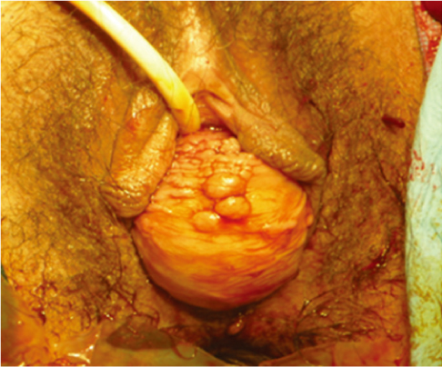
Fig. 1 A mass is protruded out through vagina in pregnancy.

Fig. 2 A protruded mass is recurrent through vagina after delivery and anterior colporrhaphy.
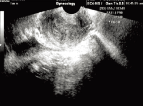
Fig. 3 Sagittal transvaginal ultrasound shows 5.1×3.1×4.0 cm sized, well defined, echogenic mass at suburethral area.
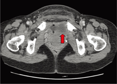
Fig. 4 Computer tomography shows 5.3×4.5 cm sized, well circumscribed mass between urethra and vagina.

Fig. 5 Histopathological finding of the vaginal tumor showing interlacing bundles of spindle smooth muscle cells, and intervening collagenous stroma without cellular atypism and mitotic figures. (A) H&E, ×12.5. (B) H&E, ×100.
Reference
-
1. Herbst AL, Mishell DR, Stenchever MA, Droegemueller W. Comprehensive gynecology. 1992. 2nd ed. St. Louis (MO): Mosby Year Book.2. Lev-Toaff AS, Coleman BG, Arger PH, Mintz MC, Arenson RL, Toaff ME. Leiomyomas in pregnancy: sonographic study. Radiology. 1987. 164:375–380.3. Kleinwaechter L. Die bindegewebigen and myomatosen Neubildugen der Vagina. Z Heilkd. 1882. 3:335–358.4. Tourneux JP. Les fibromes du vagina. Prog Med Paris. 1934. 41:1568–1570.5. Phillips J. On fibromyomata of the vagina. Br Med J. 1899. 1:262–264.6. Liu MM. Fibromyoma of the vagina. Eur J Obstet Gynecol Reprod Biol. 1988. 29:321–328.7. Douglas RG, Stromme WB, Zuspan FP, Quilligan EJ. Douglas-Stromme operative obstetrics. 1988. 5th ed. Norwalk (CT): Appleton & Lange.8. Dhaliwal LK, Das I, Gopalan S. Recurrent leiomyoma of the vagina. Int J Gynaecol Obstet. 1992. 37:281–283.9. Bernnett HG Jr, Ehrlich MM. Myoma of the vagina. Am J Obstet Gynecol. 1941. 42:314–315.10. Pulfus E, Newcomer J. Vaginal wall mass. Obstet Gynecol Surv. 1999. 54:149–150.11. O'Boyle AL, O'Boyle JD, Calhoun B, Davis GD. Pelvic organ support in pregnancy and postpartum. Int Urogynecol J Pelvic Floor Dysfunct. 2005. 16:69–72.12. Ruggieri AM, Brody JM, Curhan RP. Vaginal leiomyoma. A case report with imaging findings. J Reprod Med. 1996. 41:875–877.13. Bolt JM, Schutter EM. A tumor in the paracolpium. A case report. Eur J Obstet Gynecol Reprod Biol. 1998. 76:233–236.14. Gorlin RJ, Koutlas IG. Multiple schwannomas, multiple nevi, and multiple vaginal leiomyomas: a new dominant syndrome. Am J Med Genet. 1998. 78:76–81.
