Korean J Obstet Gynecol.
2011 Jan;54(1):1-10. 10.5468/KJOG.2011.54.1.1.
New aspect in management of fetal growth restriction
- Affiliations
-
- 1Department of Obstetrics and Gynecology, Chonnam National University Medical School, Gwangju, Korea. jwkimmd@jnu.ac.kr
- KMID: 2274016
- DOI: http://doi.org/10.5468/KJOG.2011.54.1.1
Abstract
- Fetal growth restriction (FGR) is one of the most common and complex diseases and it is associated with increased perinatal mortality and morbidity. FGR influences the long-term health of neonates and their offspring. The aim of obstetric management is to identify growth-restricted fetuses at risk of severe intrauterine hypoxia, to monitor their health and to deliver when the adverse outcome is imminent. This review aims to investigate the new aspects of FGR including the diagnosis, antenatal surveillance, and clinical management.
MeSH Terms
Figure
-
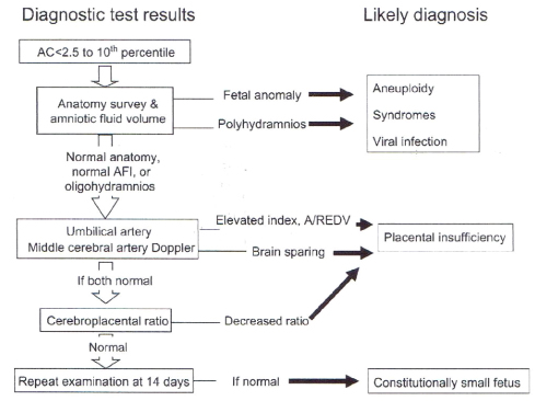
Fig. 1 Diagnostic approach to the small fetus with a decreased abdominal circumference. AC, abdominal circumference; AFI, amniotic fluid index; A/REDV, absent/reversed end-diastolic velocity (Baschat AA. J Perinat Med 2010; 38: 239-46, with permission from du Gruyter).
Reference
-
1. Ounsted M, Moar V, Scott WA. Perinatal morbidity and mortality in small-for-dates babies: the relative importance of some maternal factors. Early Hum Dev. 1981. 5:367–375.2. Rasmussen S, Irgens LM, Dalaker K. A history of placental dysfunction and risk of placental abruption. Paediatr Perinat Epidemiol. 1999. 13:9–21.3. Zhang J, Merialdi M, Platt LD, Kramer MS. Defining normal and abnormal fetal growth: promises and challenges. Am J Obstet Gynecol. 2010. 202:522–528.4. Lindqvist PG, Molin J. Does antenatal identification of small-for-gestational age fetuses significantly improve their outcome? Ultrasound Obstet Gynecol. 2005. 25:258–264.5. Mahsud-Dornan S, Dornan JC. IUGR: a contemporary review. Best Pract Res Clin Obstet Gynaecol. 2009. 23:739–740.6. Wisser J, Dirschedl P, Krone S. Estimation of gestational age by transvaginal sonographic measurement of greatest embryonic length in dated human embryos. Ultrasound Obstet Gynecol. 1994. 4:457–462.7. Ott WJ. Sonographic diagnosis of fetal growth restriction. Clin Obstet Gynecol. 2006. 49:295–307.8. Jones TB, Wolfe HM, Zador IE. Biparietal diameter and femur length discrepancies: are maternal characteristics important? Ultrasound Obstet Gynecol. 1991. 1:405–409.9. Shepard MJ, Richards VA, Berkowitz RL, Warsof SL, Hobbins JC. An evaluation of two equations for predicting fetal weight by ultrasound. Am J Obstet Gynecol. 1982. 142:47–54.10. Hadlock FP, Harrist RB, Carpenter RJ, Deter RL, Park SK. Sonographic estimation of fetal weight. The value of femur length in addition to head and abdomen measurements. Radiology. 1984. 150:535–540.11. Bobrow CS, Soothill PW. Fetal growth velocity: a cautionary tale. Lancet. 1999. 353:1460.12. Smith GC, Smith MF, McNay MB, Fleming JE. The relation between fetal abdominal circumference and birthweight: findings in 3512 pregnancies. Br J Obstet Gynaecol. 1997. 104:186–190.13. Abramowicz JS, Robischon K, Cox C. Incorporating sonographic cheek-to-cheek diameter, biparietal diameter and abdominal circumference improves weight estimation in the macrosomic fetus. Ultrasound Obstet Gynecol. 1997. 9:409–413.14. Gardeil F, Greene R, Stuart B, Turner MJ. Subcutaneous fat in the fetal abdomen as a predictor of growth restriction. Obstet Gynecol. 1999. 94:209–212.15. Larciprete G, Di Pierro G, Barbati G, Deaibess T, Jarvis S, Valensise H, et al. Could birthweight prediction models be improved by adding fetal subcutaneous tissue thickness? J Obstet Gynaecol Res. 2008. 34:18–26.16. Mintz MC, Landon MB, Gabbe SG, Marinelli DL, Ludmir J, Grumbach K, et al. Shoulder soft tissue width as a predictor of macrosomia in diabetic pregnancies. Am J Perinatol. 1989. 6:240–243.17. Sood AK, Yancey M, Richards D. Prediction of fetal macrosomia using humeral soft tissue thickness. Obstet Gynecol. 1995. 85:937–940.18. Lee W, Balasubramaniam M, Deter RL, Yeo L, Hassan SS, Gotsch F, et al. New fetal weight estimation models using fractional limb volume. Ultrasound Obstet Gynecol. 2009. 34:556–565.19. Lee W, Comstock CH, Kirk JS, Smith RS, Monck JW, Deenadayalu R, et al. Birthweight prediction by three-dimensional ultrasonographic volumes of the fetal thigh and abdomen. J Ultrasound Med. 1997. 16:799–805.20. Song TB, Moore TR, Lee JI, Kim YH, Kim EK. Fetal weight prediction by thigh volume measurement with three-dimensional ultrasonography. Obstet Gynecol. 2000. 96:157–161.21. Lubchenco LO, Hansman C, Dressler M, Boyd E. Intrauterine Growth as Estimated from Liveborn Birth-Weight Data at 24 to 42 Weeks of Gestation. Pediatrics. 1963. 32:793–800.22. Baschat AA, Weiner CP. Umbilical artery doppler screening for detection of the small fetus in need of antepartum surveillance. Am J Obstet Gynecol. 2000. 182:154–158.23. Ott WJ. Intrauterine growth restriction and Doppler ultrasonography. J Ultrasound Med. 2000. 19:661–665.24. Baschat AA. Integrated fetal testing in growth restriction: combining multivessel Doppler and biophysical parameters. Ultrasound Obstet Gynecol. 2003. 21:1–8.25. Ott WJ. Diagnosis of intrauterine growth restriction: comparison of ultrasound parameters. Am J Perinatol. 2002. 19:133–137.26. Baschat AA, Hecher K. Fetal growth restriction due to placental disease. Semin Perinatol. 2004. 28:67–80.27. Salafia CM, Minior VK, Pezzullo JC, Popek EJ, Rosenkrantz TS, Vintzileos AM. Intrauterine growth restriction in infants of less than thirty-two weeks' gestation: associated placental pathologic features. Am J Obstet Gynecol. 1995. 173:1049–1057.28. Baschat AA, Gembruch U, Harman CR. The sequence of changes in Doppler and biophysical parameters as severe fetal growth restriction worsens. Ultrasound Obstet Gynecol. 2001. 18:571–577.29. Harman CR, Baschat AA. Comprehensive assessment of fetal wellbeing: which Doppler tests should be performed? Curr Opin Obstet Gynecol. 2003. 15:147–157.30. Bukowski R, Uchida T, Smith GC, Malone FD, Ball RH, Nyberg DA, et al. Individualized norms of optimal fetal growth: fetal growth potential. Obstet Gynecol. 2008. 111:1065–1076.31. Gagnon A, Wilson RD, Audibert F, Allen VM, Blight C, Brock JA, et al. Obstetrical complications associated with abnormal maternal serum markers analytes. J Obstet Gynaecol Can. 2008. 30:918–949.32. Maulik D, Frances Evans J, Ragolia L. Fetal growth restriction: pathogenic mechanisms. Clin Obstet Gynecol. 2006. 49:219–227.33. Chaiworapongsa T, Espinoza J, Gotsch F, Kim YM, Kim GJ, Goncalves LF, et al. The maternal plasma soluble vascular endothelial growth factor receptor-1 concentration is elevated in SGA and the magnitude of the increase relates to Doppler abnormalities in the maternal and fetal circulation. J Matern Fetal Neonatal Med. 2008. 21:25–40.34. Crispi F, Dominguez C, Llurba E, Martin-Gallan P, Cabero L, Gratacos E. Placental angiogenic growth factors and uterine artery Doppler findings for characterization of different subsets in preeclampsia and in isolated intrauterine growth restriction. Am J Obstet Gynecol. 2006. 195:201–207.35. Romero R, Nien JK, Espinoza J, Todem D, Fu W, Chung H, et al. A longitudinal study of angiogenic (placental growth factor) and anti-angiogenic (soluble endoglin and soluble vascular endothelial growth factor receptor-1) factors in normal pregnancy and patients destined to develop preeclampsia and deliver a small for gestational age neonate. J Matern Fetal Neonatal Med. 2008. 21:9–23.36. Dunger DB, Petry CJ, Ong KK. Genetic variations and normal fetal growth. Horm Res. 2006. 65:Suppl 3. 34–40.37. Kaku K, Osada H, Seki K, Sekiya S. Insulin-like growth factor 2 (IGF2) and IGF2 receptor gene variants are associated with fetal growth. Acta Paediatr. 2007. 96:363–367.38. Baschat AA. Fetal growth restriction-from observation to intervention. J Perinat Med. 2010. 38:239–246.39. FitzGerald DE, Drumm JE. Non-invasive measurement of human fetal circulation using ultrasound: a new method. Br Med J. 1977. 2:1450–1451.40. Trudinger BJ, Giles WB, Cook CM, Bombardieri J, Collins L. Fetal umbilical artery flow velocity waveforms and placental resistance: clinical significance. Br J Obstet Gynaecol. 1985. 92:23–30.41. Karsdorp VH, van Vugt JM, van Geijn HP, Kostense PJ, Arduini D, Montenegro N, et al. Clinical significance of absent or reversed end diastolic velocity waveforms in umbilical artery. Lancet. 1994. 344:1664–1668.42. Brodszki J, Hernandez-Andrade E, Gudmundsson S, Dubiel M, Mandruzzato GP, Laurini R, et al. Can the degree of retrograde diastolic flow in abnormal umbilical artery flow velocity waveforms predict pregnancy outcome? Ultrasound Obstet Gynecol. 2002. 19:229–234.43. Thornton JG, Hornbuckle J, Vail A, Spiegelhalter DJ, Levene M. Infant wellbeing at 2 years of age in the Growth Restriction Intervention Trial (GRIT): multicentred randomised controlled trial. Lancet. 2004. 364:513–520.44. Alfirevic Z, Neilson JP. WITHDRAWN. Doppler ultrasound for fetal assessment in high risk pregnancies. Cochrane Database Syst Rev. 2010. (1):CD000073.45. Goffinet F, Paris-Llado J, Nisand I, Breart G. Umbilical artery Doppler velocimetry in unselected and low risk pregnancies: a review of randomised controlled trials. Br J Obstet Gynaecol. 1997. 104:425–430.46. Harrington K, Cooper D, Lees C, Hecher K, Campbell S. Doppler ultrasound of the uterine arteries: the importance of bilateral notching in the prediction of pre-eclampsia, placental abruption or delivery of a small-for-gestational-age baby. Ultrasound Obstet Gynecol. 1996. 7:182–188.47. Bricker L, Neilson JP. Routine doppler ultrasound in pregnancy. Cochrane Database Syst Rev. 2000. (2):CD001450.48. Vergani P, Roncaglia N, Andreotti C, Arreghini A, Teruzzi M, Pezzullo JC, et al. Prognostic value of uterine artery Doppler velocimetry in growth-restricted fetuses delivered near term. Am J Obstet Gynecol. 2002. 187:932–936.49. Sebire NJ. Umbilical artery Doppler revisited: pathophysiology of changes in intrauterine growth restriction revealed. Ultrasound Obstet Gynecol. 2003. 21:419–422.50. Mari G. Doppler ultrasonography in obstetrics: from the diagnosis of fetal anemia to the treatment of intrauterine growth-restricted fetuses. Am J Obstet Gynecol. 2009. 200:613.e1–613.e9.51. Hershkovitz R, Kingdom JC, Geary M, Rodeck CH. Fetal cerebral blood flow redistribution in late gestation: identification of compromise in small fetuses with normal umbilical artery Doppler. Ultrasound Obstet Gynecol. 2000. 15:209–212.52. Severi FM, Bocchi C, Visentin A, Falco P, Cobellis L, Florio P, et al. Uterine and fetal cerebral Doppler predict the outcome of third-trimester small-for-gestational age fetuses with normal umbilical artery Doppler. Ultrasound Obstet Gynecol. 2002. 19:225–228.53. To WW, Chan AM, Mok KM. Use of umbilical-cerebral Doppler ratios in predicting fetal growth restriction in near-term fetuses. Aust N Z J Obstet Gynaecol. 2005. 45:130–136.54. Scherjon SA, Oosting H, Smolders-DeHaas H, Zondervan HA, Kok JH. Neurodevelopmental outcome at three years of age after fetal 'brain-sparing'. Early Hum Dev. 1998. 52:67–79.55. Cruz-Martinez R, Figueras F, Oros D, Padilla N, Meler E, Hernandez-Andrade E, et al. Cerebral blood perfusion and neurobehavioral performance in full-term small-for-gestational-age fetuses. Am J Obstet Gynecol. 2009. 201:474.e1–474.e7.56. Eixarch E, Meler E, Iraola A, Illa M, Crispi F, Hernandez-Andrade E, et al. Neurodevelopmental outcome in 2-year-old infants who were small-for-gestational age term fetuses with cerebral blood flow redistribution. Ultrasound Obstet Gynecol. 2008. 32:894–899.57. Borrell A, Antolin E, Costa D, Farre MT, Martinez JM, Fortuny A. Abnormal ductus venosus blood flow in trisomy 21 fetuses during early pregnancy. Am J Obstet Gynecol. 1998. 179:1612–1617.58. Baschat AA, Gembruch U, Weiner CP, Harman CR. Qualitative venous Doppler waveform analysis improves prediction of critical perinatal outcomes in premature growth-restricted fetuses. Ultrasound Obstet Gynecol. 2003. 22:240–245.59. Schwarze A, Gembruch U, Krapp M, Katalinic A, Germer U, Axt-Fliedner R. Qualitative venous Doppler flow waveform analysis in preterm intrauterine growth-restricted fetuses with ARED flow in the umbilical artery-correlation with short-term outcome. Ultrasound Obstet Gynecol. 2005. 25:573–579.60. Hecher K, Bilardo CM, Stigter RH, Ville Y, Hackeloer BJ, Kok HJ, et al. Monitoring of fetuses with intrauterine growth restriction: a longitudinal study. Ultrasound Obstet Gynecol. 2001. 18:564–570.61. Ferrazzi E, Bozzo M, Rigano S, Bellotti M, Morabito A, Pardi G, et al. Temporal sequence of abnormal Doppler changes in the peripheral and central circulatory systems of the severely growth-restricted fetus. Ultrasound Obstet Gynecol. 2002. 19:140–146.62. Makikallio K, Vuolteenaho O, Jouppila P, Rasanen J. Ultrasonographic and biochemical markers of human fetal cardiac dysfunction in placental insufficiency. Circulation. 2002. 105:2058–2063.63. Rizzo G, Arduini D, Romanini C. Doppler echocardiographic assessment of fetal cardiac function. Ultrasound Obstet Gynecol. 1992. 2:434–445.64. Rizzo G, Capponi A, Rinaldo D, Arduini D, Romanini C. Ventricular ejection force in growth-retarded fetuses. Ultrasound Obstet Gynecol. 1995. 5:247–255.65. Cruz-Martinez R, Figueras F, Benavides-Serralde A, Crispi F, Hernandez-Andrade E, Gratacos E. Sequence of changes in myocardial performance index in relation with aortic isthmus and ductus venosus Doppler in fetuses with early-onset intrauterine growth restriction. Ultrasound Obstet Gynecol. 2010. 12. 10. [Epub]. DOI: 10.1002/uog.8903.66. Turan OM, Turan S, Gungor S, Berg C, Moyano D, Gembruch U, et al. Progression of Doppler abnormalities in intrauterine growth restriction. Ultrasound Obstet Gynecol. 2008. 32:160–167.67. Phelan JP, Ahn MO, Smith CV, Rutherford SE, Anderson E. Amniotic fluid index measurements during pregnancy. J Reprod Med. 1987. 32:601–604.68. Chauhan SP, Sanderson M, Hendrix NW, Magann EF, Devoe LD. Perinatal outcome and amniotic fluid index in the antepartum and intrapartum periods: A meta-analysis. Am J Obstet Gynecol. 1999. 181:1473–1478.69. Gulmezoglu AM, Hofmeyr GJ. Bed rest in hospital for suspected impaired fetal growth. Cochrane Database Syst Rev. 2000. (2):CD000034.70. Say L, Gulmezoglu AM, Hofmeyr GJ. Maternal nutrient supplementation for suspected impaired fetal growth. Cochrane Database Syst Rev. 2003. (1):CD000148.71. Say L, Gulmezoglu AM, Hofmeyr GJ. Maternal oxygen administration for suspected impaired fetal growth. Cochrane Database Syst Rev. 2003. (1):CD000137.72. Gulmezoglu AM, Hofmeyr GJ. Calcium channel blockers for potential impaired fetal growth. Cochrane Database Syst Rev. 2000. (2):CD000049.73. Bernstein IM, Horbar JD, Badger GJ, Ohlsson A, Golan A. The Vermont Oxford Network. Morbidity and mortality among very-low-birth-weight neonates with intrauterine growth restriction. Am J Obstet Gynecol. 2000. 182:198–206.74. Baschat AA, Cosmi E, Bilardo CM, Wolf H, Berg C, Rigano S, et al. Predictors of neonatal outcome in early-onset placental dysfunction. Obstet Gynecol. 2007. 109:253–261.75. Brodszki J, Morsing E, Malcus P, Thuring A, Ley D, Marsal K. Early intervention in management of very preterm growth-restricted fetuses: 2-year outcome of infants delivered on fetal indication before 30 gestational weeks. Ultrasound Obstet Gynecol. 2009. 34:288–296.76. Marsal K. Obstetric management of intrauterine growth restriction. Best Pract Res Clin Obstet Gynaecol. 2009. 23:857–870.77. Froen JF, Gardosi JO, Thurmann A, Francis A, Stray-Pedersen B. Restricted fetal growth in sudden intrauterine unexplained death. Acta Obstet Gynecol Scand. 2004. 83:801–807.78. Baschat AA, Berg C, Turan O, Turan S, Galan H, Thilaganathan B, et al. Natural history of stillbirth in placenta based fetal growth restriction-implications for surveillance. Annual Meeting of the Society for Maternal-Fetal Medicine. Am J Obstet Gynecol. 2008. 199:S198.79. Bahado-Singh RO, Kovanci E, Jeffres A, Oz U, Deren O, Copel J, et al. The Doppler cerebroplacental ratio and perinatal outcome in intrauterine growth restriction. Am J Obstet Gynecol. 1999. 180:750–756.80. Hecher K, Spernol R, Stettner H, Szalay S. Potential for diagnosing imminent risk to appropriate- and small-for-gestational-age fetuses by Doppler sonographic examination of umbilical and cerebral arterial blood flow. Ultrasound Obstet Gynecol. 1992. 2:266–271.81. Kaukola T, Rasanen J, Herva R, Patel DD, Hallman M. Suboptimal neurodevelopment in very preterm infants is related to fetal cardiovascular compromise in placental insufficiency. Am J Obstet Gynecol. 2005. 193:414–420.82. Baschat AA, Viscardi RM, Hussey-Gardner B, Hashmi N, Harman C. Infant neurodevelopment following fetal growth restriction: relationship with antepartum surveillance parameters. Ultrasound Obstet Gynecol. 2009. 33:44–50.83. Roza SJ, Steegers EA, Verburg BO, Jaddoe VW, Moll HA, Hofman A, et al. What is spared by fetal brain-sparing? Fetal circulatory redistribution and behavioral problems in the general population. Am J Epidemiol. 2008. 168:1145–1152.
- Full Text Links
-
- Actions
-
Cited
- CITED
-
- Close
- Share
-
- Similar articles
-
- The Evaluation and Management of Fetal Growth Restriction
- Recent advances in management of fetal growth restriction
- Neonatal Management and Outcome of Fetal Growth Restriction
- Relationships between TNF-alpha and fetal growth restriction in preeclamptic women and normotensive pregnancies
- Diagnosis and Management of Fetal Growth Restriction
