Mature cystic teratoma in a 5-year-old girl presenting as urinary frequency: A case report
- Affiliations
-
- 1Department of Obstetrics and Gynecology, Seoul Medical Center, Seoul, Korea. harmony4@catholic.ac.kr
- KMID: 2273947
- DOI: http://doi.org/10.5468/kjog.2010.53.7.652
Abstract
- Ovarian tumors are rare in children. Their incidence is estimated to be about 2.6 cases per 100,000 girls per year. About 1/3 of all childhood ovarian tumors are reported to be malignant. Germ cell tumors are more frequent than epithelial and sex cord stromal tumors in children and teratoma is the most common germ cell tumor occurring in children. In most cases, the presenting symptoms in childhood included abdominal pain, an abdominal mass, abdominal distention and so on. These non-specific symptoms and low incidence lead to suspicions of more common diseases, so the diagnosis of ovarian masses in childhood is difficult. We experienced a rare case of mature cystic teratoma in a 5-year-old girl with urinary frequency without abdominal discomfort despite the large size. The pre-operative magnetic resonance imaging finding showed unusual characteristics, rising suspicion of malignancy. So, we present this case with a brief review of literature.
MeSH Terms
Figure
-
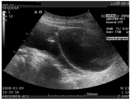
Fig. 1 Transabdominal ultrasonogram shows huge multiple septated hypoechogenic cystic mass with some mixed echogenic area and posterior acoustic shadowing.

Fig. 2 T1 weighted axial magnetic resonance image shows the minute size of high signal intensity lesion at peripheral area (arrow head), which misleaded this fat component of the ovarian cyst for other tissue. Also, the septated thick wall (arrow) can be noted.
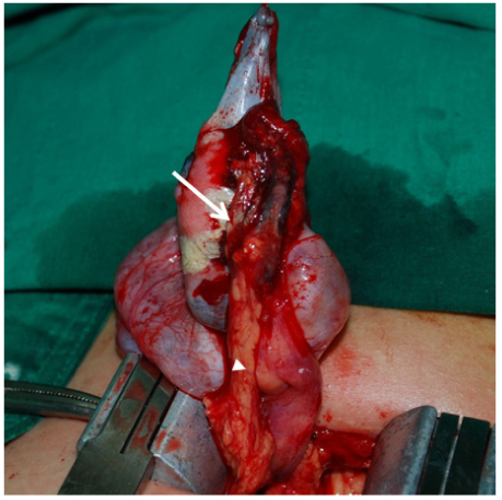
Fig. 3 Gross photography of right lobulating mass after aspiration, adhered to omentum (arrow head) and covered with hair and fat tissue (arrow).
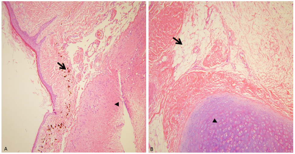
Fig. 4 Microscopic findings of mature cystic teratoma showing. (A) Mature stratified squamous epithelium with melanin pigment (arrow) and underline brain tissue (arrow head) with glial cell and neuron (H&E stain, ×100), (B) Fat cell (arrow) and cartilage (arrow head) (H&E stain, ×100).
Reference
-
1. Skinner MA, Schlatter MG, Heifetz SA, Grosfeld JL. Ovarian neoplasms in children. Arch Surg. 1993. 128:849–853.2. De Backer A, Madern GC, Oosterhuis JW, Hakvoort-Cammel FG, Hazebroek FW. Ovarian germ cell tumors in children: a clinical study of 66 patients. Pediatr Blood Cancer. 2006. 46:459–464.3. Caruso PA, Marsh MR, Minkowitz S, Karten G. An intense clinicopathologic study of 305 teratomas of the ovary. Cancer. 1971. 27:343–348.4. van Winter JT, Simmons PS, Podratz KC. Surgically treated adnexal masses in infancy, childhood, and adolescence. Am J Obstet Gynecol. 1994. 170:1780–1786.5. Lucraft HH. Ovarian tumours in children-a review of 40 cases. Clin Radiol. 1979. 30:279–285.6. Abell MR, Johnson VJ, Holtz F. Ovarian neoplasms in childhood and adolescence. I. Tumors of germ cell origin. Am J Obstet Gynecol. 1965. 92:1059–1081.7. Norris HJ, Jensen RD. Relative frequency of ovarian neoplasms in children and adolescents. Cancer. 1972. 30:713–719.8. Shim SI, Hur SY, Lee GS, Kim SJ, Kim EJ, Song SK, et al. Clinicopathological observation on ovarian tumors in premenarcheal years. Korean J Obstet Gynecol. 1998. 41:2072–2079.9. Park SH, Park BJ, Kim YW, Kim TE, Maeng LS. Ruptured ovarian mature cystic teratoma with jaw bone and teeth: a case report. Korean J Obstet Gynecol. 2008. 51:1571–1575.10. Ein SH, Darte JM, Stephens CA. Cystic and solid ovarian tumors in children: a 44-year review. J Pediatr Surg. 1970. 5:148–156.11. Adams Hillard PJ. Berek JS, Novak E, editors. Benign disease of the female reproductive tract. Berek & Novak's gynecology. 2007. 14th ed. Philadelphia: Lippincott Williams & Wilkins;441.12. Laing FC, Van Dalsem VF, Marks WM, Barton JL, Martinez DA. Dermoid cysts of the ovary: their ultrasonographic appearances. Obstet Gynecol. 1981. 57:99–104.13. Friedman AC, Pyatt RS, Hartman DS, Downey EF Jr, Olson WB. CT of benign cystic teratomas. AJR Am J Roentgenol. 1982. 138:659–665.14. Bouic-Pages E, Perrochia H, Merigeaud S, Giacalone PY, Taourel P. MR Imaging of primary ovarian tumors with pathologic correlation. J Radiol. 2009. 90:787–802.15. Dede M, Gungor S, Yenen MC, Alanbay I, Duru NK, Hasimi A. CA19-9 may have clinical significance in mature cystic teratomas of the ovary. Int J Gynecol Cancer. 2006. 16:189–193.
- Full Text Links
-
- Actions
-
Cited
- CITED
-
- Close
- Share
-
- Similar articles
-
- Squamous Cell Carcinoma, Adenocarcinoma and Papillary Cell Carcinoma Arising from the Mature Cystic Teratoma of the Ovary: Case Report
- A Case of Mature cystic teratoma in Omentum
- A Case of Papillary Carcinoma of Thyroid Gland Arising from Ovarian Mature Cystic Teratoma
- A Case of Anaplastic Carcinoma Arising from Mature Cystic Teratoma of Ovary
- Congenital Hydrocolpos Mimicking a Mature Cystic Teratoma in the Pelvis
