Torque control during lingual anterior retraction without posterior appliances
- Affiliations
-
- 1Division of Orthodontics, Department of Dentistry, The Catholic University of Korea, College of Medicine, Seoul, Korea.
- 2Department of Orthodontics, School of Dentistry, Kyung Hee University, Seoul, Korea. bravortho@hanmail.net
- 3Department of Orthodontics, Asan Medical Center, University of Ulsan College of Medicine, Seoul, Korea.
- 4Division of Orthodontics, Department of Dentistry, Ajou University College of Medicine, Suwon, Korea.
- 5Department of Dentistry, Ewha Womans University School of Medicine, Seoul, Korea.
- 6University of California, San Francisco, San Francisco, CA, USA.
- KMID: 2273463
- DOI: http://doi.org/10.4041/kjod.2013.43.1.3
Abstract
OBJECTIVE
To evaluate the factors that affect torque control during anterior retraction when utilizing the C-retractor with a palatal miniplate as an exclusive source of anchorage without posterior appliances.
METHODS
The C-retractor was modeled using a 3-dimensional beam element (0.9-mm-diameter stainless-steel wire) attached to mesh bonding pads. Various vertical heights and 2 attachment positions for the lingual anterior retraction hooks (LARHs) were evaluated. A force of 200 g was applied from each side hook of the miniplate to the splinted segment of 6 or 8 anterior teeth.
RESULTS
During anterior retraction, an increase in the LARH vertical height increased the amount of lingual root torque and intrusion of the incisors. In particular, with increasing vertical height, the tooth displacement pattern changed from controlled tipping to bodily displacement and then to lingual root displacement. The effects were enhanced when the LARH was located between the central and lateral incisors, as compared to when the LARH was located between the lateral incisors and canines.
CONCLUSIONS
Three-dimensional lingual anterior retraction of the 6 or 8 anterior teeth can be accomplished using the palatal miniplate as the only anchorage source. Using LARHs at different heights or positions affects the quality of torque and intrusion.
Keyword
Figure
-
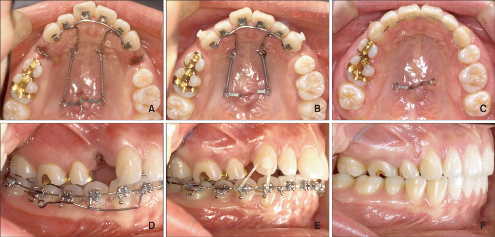
Figure 1 A first premolar extraction case using the lingual biocreative therapy. A and D, Lingual en masse retraction forces are initiated. B and E, Seven months of en masse retraction. Triangular elastics were applied to the canine for vertical control. C and F, Post-treatment. The total treatment period was 13 months.

Figure 2 A second premolar extraction case using the lingual biocreative therapy. A to C, Pre-treatment photos show dens evaginatus on #15 and a malformed #25. D to F, One month after en masse retraction force is initiated. G to I, Four months of en masse retraction.
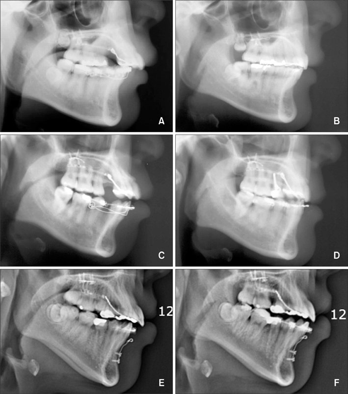
Figure 3 Pre- and post-lateral cephalograms. A and B, Low lingual anterior retraction hook (LARH) - the patient needed controlled lingual tipping; hence, a LARH vertical height of 4 mm was used. C and D, High LARH - the patient needed bodily tooth movement; thus, a LARH vertical height of 13 mm was used. E and F, Second premolars were extracted due to internal resorption, so the 8 anterior teeth were retracted using the lingual biocreative therapy.
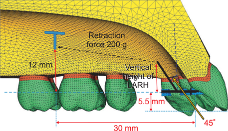
Figure 4 Three-dimensional finite element mesh with teeth, periodontal ligament, alveolar bone of the maxillary dentition, and C-retractor with the low lingual anterior retraction hook (LARH).
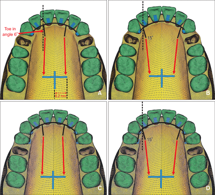
Figure 5 Schematic representation of the coordinate system of the lingual biocreative therapy with the low lingual anterior retraction hook (LARH) at different positions. A, Condition 1: the LARHs, made of 0.9-mm round stainless steel, were placed between the upper central and lateral incisors with 6° of toe in angle. B, Condition 2: the LARHs were placed between the lateral incisors and canines with 15° of toe in angle. C, Condition 3: the LARHs were placed between the upper central and lateral incisors after second premolar extraction. D, Condition 4: the LARHs were placed between the lateral incisors and canines with 15° of toe in angle after second premolar extraction.

Figure 6 Comparison of the effects of the different lengths and positions of the low lingual anterior retraction hooks (LARHs) in the C-retractor in the three dimensional finite element model. Tooth axies graph (incisor, midpoint of incisal edge to root apex; canine, cusp tip to root apex) magnified tooth displacement 70 times. Solid line means before displacement and a dotted line means after displacement (circles, central incisor; squares, lateral incisor; canine, triangles). Condition 1, The LARHs were placed between the upper central and lateral incisors after first premolar extraction. Condition 2, The LARHs were placed between the upper lateral incisors and canines after first premolar extraction. Condition 3, The LARHs were placed between the upper central and lateral incisors after second premolar extraction. Condition 4, The LARHs were placed between the upper lateral incisors and canines after second premolar extraction.

Figure 7 Comparison of the vertical effects (Z-axis) of the different heights and positions of the low lingual anterior retraction hooks (LARHs) in the three dimensional finite element model.
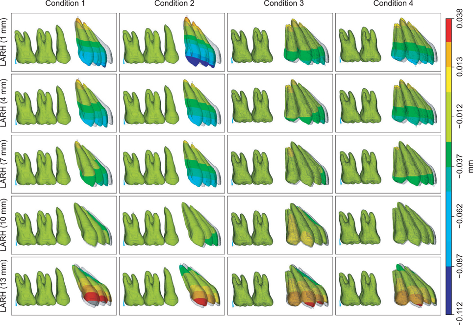
Figure 8 Comparison of the sagittal effects (Y-axis) of the different heights and positions of the low lingual anterior retraction hooks (LARHs) in the three dimensional finite element model.

Figure 9 A, When the lingual anterior retraction hook is located distal to the central incisors (condition 1 or 3). B, Part of the extraction space is closed by C-retractor. C, The canine can be segmented from the C-retractor for detailing. D, Incisors and canines are decrowded with conventional bracket system.
Cited by 5 articles
-
Displacement pattern of the anterior segment using antero-posterior lingual retractor combined with a palatal plate
Kyung-Won Seo, Soon-Yong Kwon, Kyung A Kim, Ki-Ho Park, Seong-Hun Kim, Hyo-Won Ahn, Gerald Nelson
Korean J Orthod. 2015;45(6):289-298. doi: 10.4041/kjod.2015.45.6.289.Effect of labiolingual inclination of a maxillary central incisor and surrounding alveolar bone loss on periodontal stress: A finite element analysis
Sung-Hwan Choi, Young-Hoon Kim, Kee-Joon Lee, Chung-Ju Hwang
Korean J Orthod. 2016;46(3):155-162. doi: 10.4041/kjod.2016.46.3.155.The effects of alveolar bone loss and miniscrew position on initial tooth displacement during intrusion of the maxillary anterior teeth: Finite element analysis
Sun-Mi Cho, Sung-Hwan Choi, Sang-Jin Sung, Hyung-Seog Yu, Chung-Ju Hwang
Korean J Orthod. 2016;46(5):310-322. doi: 10.4041/kjod.2016.46.5.310.Palatal en-masse retraction of segmented maxillary anterior teeth: A finite element study
Jae Hyun Park, Yoon-Ah Kook, Yukio Kojima, Sunock Yun, Jong-Moon Chae
Korean J Orthod. 2019;49(3):188-193. doi: 10.4041/kjod.2019.49.3.188.Type of tooth movement during en masse retraction of the maxillary anterior teeth using labial versus lingual biocreative therapy in adults: A randomized clinical trial
Mais M. Sadek, Noha E. Sabet, Islam T. Hassan
Korean J Orthod. 2019;49(6):381-392. doi: 10.4041/kjod.2019.49.6.381.
Reference
-
1. Ye L, Kula KS. Status of lingual orthodontics. World J Orthod. 2006. 7:361–368.2. Takemoto K, Scuzzo G. The straight-wire concept in lingual orthodontics. J Clin Orthod. 2001. 35:46–52.3. Gorman CJ Jr. Lingual orthodontics. Dent Clin North Am. 1997. 41:111–125.4. Hong RK, Sohn HW. Update on the Fujita lingual bracket. J Clin Orthod. 1999. 33:136–142.5. Wiechmann D, Gerss J, Stamm T, Hohoff A. Prediction of oral discomfort and dysfunction in lingual orthodontics: a preliminary report. Am J Orthod Dentofacial Orthop. 2008. 133:359–364.
Article6. Miyawaki S, Yasuhara M, Koh Y. Discomfort caused by bonded lingual orthodontic appliances in adult patients as examined by retrospective questionnaire. Am J Orthod Dentofacial Orthop. 1999. 115:83–88.
Article7. Stamm T, Hohoff A, Ehmer U. A subjective comparison of two lingual bracket systems. Eur J Orthod. 2005. 27:420–426.
Article8. Cacciafesta V, Sfondrini MF. One-appointment correction of a scissor bite with 2D lingual brackets and fiber-reinforced composites. J Clin Orthod. 2006. 40:409–411.9. Park HS. A miniscrew-assisted transpalatal arch for use in lingual orthodontics. J Clin Orthod. 2006. 40:12–16.10. Hong RK, Heo JM, Ha YK. Lever-arm and mini-implant system for anterior torque control during retraction in lingual orthodontic treatment. Angle Orthod. 2005. 75:129–141.11. Kim KH, Lee KJ, Cha JY, Park YC. Finite element analysis of effectiveness of lever arm in lingual sliding mechanics. Korean J Orthod. 2011. 41:324–336.
Article12. Chung KR, Jeong DM, Park HJ, Kim SH, Nelson G. Severe bidentoalveolar protrusion treated with lingual biocreative therapy using palatal miniplate. Korean J Orthod. 2010. 40:276–287.
Article13. Chung KR, Kook YA, Kim SH, Mo SS, Jung JA. Class II malocclusion treated by combining a lingual retractor and a palatal plate. Am J Orthod Dentofacial Orthop. 2008. 133:112–123.
Article14. Kim S, Park Y, Chung K. Severe anterior open bite malocclusion with multiple odontoma treated by C-lingual retractor and horseshoe mechanics. Angle Orthod. 2003. 73:206–212.15. Kim JS, Kim SH, Kook YA, Chung KR, Nelson G. Analysis of lingual en masse retraction combining a C-lingual retractor and a palatal plate. Angle Orthod. 2011. 81:662–669.
Article16. Andrews LF. The six keys to normal occlusion. Am J Orthod. 1972. 62:296–309.
Article17. Germane N, Bentley BE Jr, Isaacson RJ. Three biologic variables modifying faciolingual tooth angulation by straight-wire appliances. Am J Orthod Dentofacial Orthop. 1989. 96:312–319.
Article18. Coolidge ED. The thickness of the human periodontal membrane. J Am Dent Assoc. 1937. 24:1260–1270.
Article19. Tanne K, Sakuda M, Burstone CJ. Three-dimensional finite element analysis for stress in the periodontal tissue by orthodontic forces. Am J Orthod Dentofacial Orthop. 1987. 92:499–505.
Article20. Poppe M, Bourauel C, Jäger A. Determination of the elasticity parameters of the human periodontal ligament and the location of the center of resistance of single-rooted teeth a study of autopsy specimens and their conversion into finite element models. J Orofac Orthop. 2002. 63:358–370.
Article21. Kim MJ, Park SH, Kim HS, Mo SS, Sung SJ, Jang GW, et al. Effects of orthodontic mini-implant position in the dragon helix appliance on tooth displacement and stress distribution: a three-dimensional finite element analysis. Korean J Orthod. 2011. 41:191–199.
Article22. Bae SM, Park HS, Kyung HM, Kwon OW, Sung JH. Clinical application of micro-implant anchorage. J Clin Orthod. 2002. 36:298–302.23. Burstone CJ. The segmented arch approach to space closure. Am J Orthod. 1982. 82:361–378.
Article24. Sung SJ, Kim IT, Kook YA, Chun YS, Kim SH, Mo SS. Finite-element analysis of the shift in center of resistance of the maxillary dentition in relation to alveolar bone loss. Korean J Orthod. 2009. 39:278–288.
Article25. Jeong GM, Sung SJ, Lee KJ, Chun YS, Mo SS. Finite-element investigation of the center of resistance of the maxillary dentition. Korean J Orthod. 2009. 39:83–94.
Article26. Jang HJ, Roh WJ, Joo BH, Park KH, Kim SJ, Park YG. Locating the center of resistance of maxillary anterior teeth retracted by Double J Retractor with palatal miniscrews. Angle Orthod. 2010. 80:1023–1028.
Article27. Park YC, Choi YJ, Choi NC, Lee JS. Esthetic segmental retraction of maxillary anterior teeth with a palatal appliance and orthodontic mini-implants. Am J Orthod Dentofacial Orthop. 2007. 131:537–544.
Article28. Kim SH, Hwang YS, Ferreira A, Chung KR. Analysis of temporary skeletal anchorage devices used for enmasse retraction: a preliminary study. Am J Orthod Dentofacial Orthop. 2009. 136:268–276.
Article29. Mo SS, Kim SH, Sung SJ, Chung KR, Chun YS, Kook YA, et al. Factors controlling anterior torque during C-implant-dependent en-masse retraction without posterior appliances. Am J Orthod Dentofacial Orthop. 2011. 140:72–80.
Article30. Kim HS, Lee YJ, Park YG, Chung KR, Kang YG, Choo H, et al. Histologic assessment of the biological effects after speedy surgical orthodontics in a beagle animal model: a preliminary study. Korean J Orthod. 2011. 41:361–370.
Article
- Full Text Links
-
- Actions
-
Cited
- CITED
-
- Close
- Share
-
- Similar articles
-
- Incisor inclination indicator for anterior torque control during retraction in lingual orthodontic treatment
- Displacement pattern of the anterior segment using antero-posterior lingual retractor combined with a palatal plate
- Comparison of inclination and vertical changes between single-wire and double-wire retraction techniques in lingual orthodontics
- Type of tooth movement during en masse retraction of the maxillary anterior teeth using labial versus lingual biocreative therapy in adults: A randomized clinical trial
- Finite element analysis of the effects of different archwire forms and power arm positions on maxillary incisors in en masse retraction using fixed lingual orthodontic appliances
