Orthodontic and surgical management of cleidocranial dysplasia
- Affiliations
-
- 1Orthodontics Advanced Specialty Program, Ostrow School of Dentistry, University of Southern California, CA, USA.
- 2School of Dentistry, University of California, San Francisco, CA, USA.
- 3Orofacial Sciences, University of California, San Francisco, CA, USA. sneha.oberoi@ucsf.edu
- KMID: 2273417
- DOI: http://doi.org/10.4041/kjod.2013.43.5.248
Abstract
- Cleidocranial dysplasia (CCD), an autosomal dominant disorder with a prevalence of 1 in 1,000,000 individuals, is mainly caused by mutations in Runx2, a gene required for osteoblastic differentiation. It is generally characterized by hypoplastic clavicles, narrow thorax, and delayed or absent fontanel closure. Importantly, its orofacial manifestations, including midfacial hypoplasia, retained primary teeth, and impacted permanent and supernumerary teeth, severely impede the well-being of affected individuals. Successful treatment of the orofacial problems requires the combined efforts of dental specialists. However, only a few successfully treated cases have been reported because of the rarity of CCD and complexity of the treatment. This article presents the University of California, San Francisco (UCSF) treatment protocol for the dentofacial manifestations of CCD based on two treated and 17 diagnosed cases. The records of two patients with CCD who had been treated at the UCSF School of Dentistry and the treatment options reported in the literature were reviewed. The UCSF treatment protocol produced a successful case and a partially successful one (inadequate oral hygiene in the retention stage resulted in decay and loss of teeth). It provides general guidelines for successfully treating the orofacial manifestations of CCD.
MeSH Terms
Figure
-
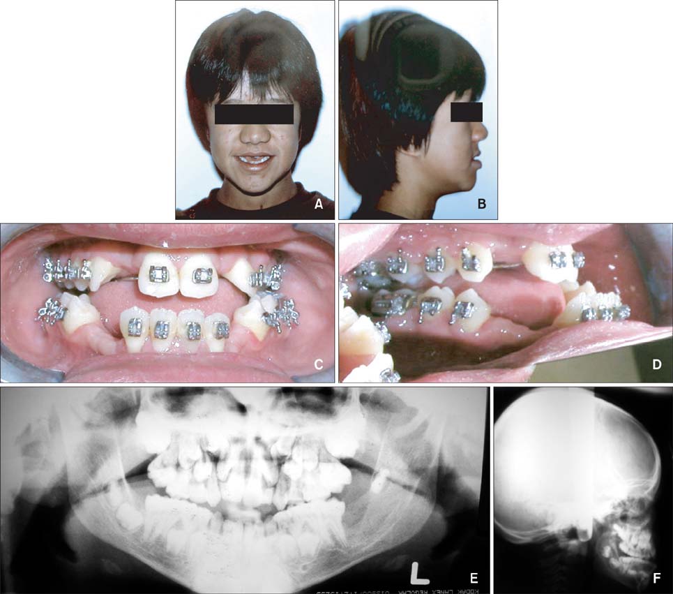
Figure 1 Initial record of patient 1 at age 10 years and 11 months. A, Frontal facial view; B, lateral facial view; C, frontal intraoral view; D, lateral intraoral view; E, panoramic radiograph; and F, lateral cephalogram.
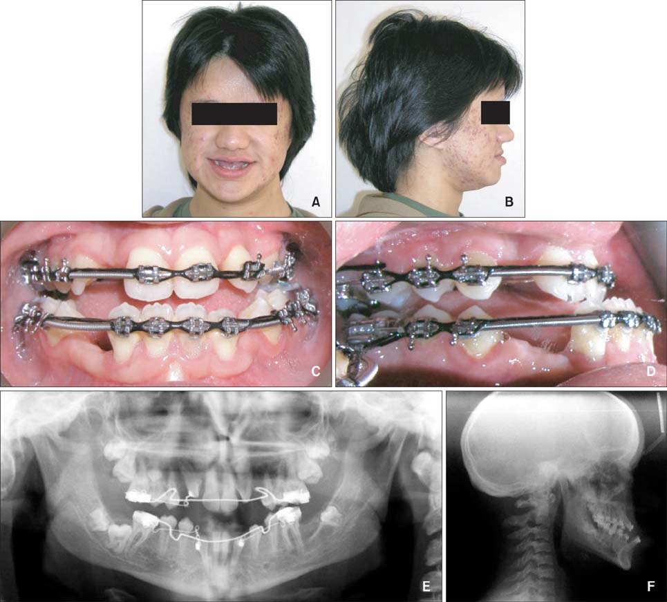
Figure 2 Progress record of patient 1 at age 17 years. A, Frontal facial view; B, lateral facial view; C, frontal intraoral view; D, lateral intraoral view; E, panoramic radiograph; and F, lateral cephalogram.

Figure 3 Progress record of patient 1 at age 23 years. A, Frontal facial view; B, lateral facial view; C, frontal intraoral view; D, lateral intraoral view; E, panoramic radiograph; and F, lateral cephalogram.

Figure 4 Final record of patient 1 at age 24 years. A, Frontal facial view; B, lateral facial view; C, frontal intraoral view; D, lateral intraoral view.
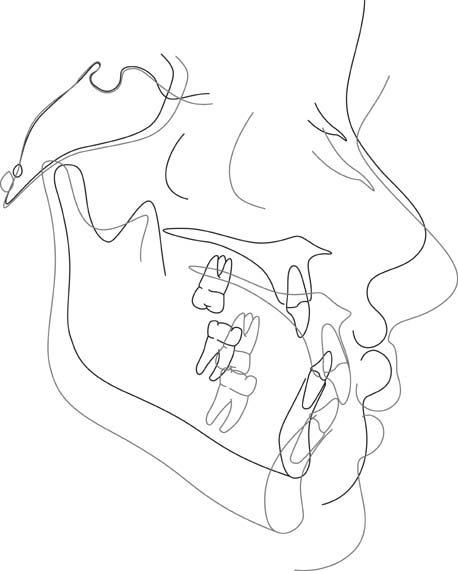
Figure 5 Superimposition of the pretreatment (dotted line) and posttreatment (solid line) lateral cephalograms in case 1.
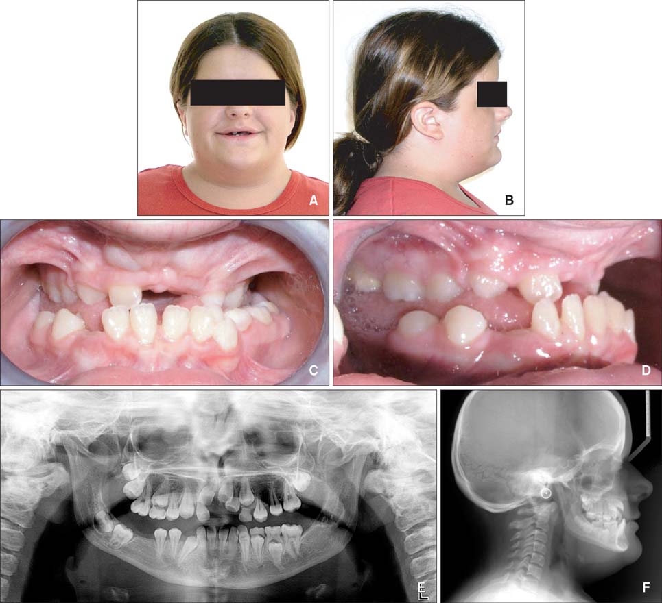
Figure 6 Initial record of patient 2 at age 14 years. A, Frontal facial view; B, lateral facial view; C, frontal intraoral view; D, lateral intraoral view; E, panoramic radiograph; and F, lateral cephalogram.
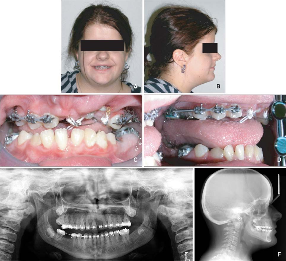
Figure 7 Progress record of patient 2 at age 17 years (A and B), 15 years (C and D), and 16 years (E and F). A, Frontal facial view; B, lateral facial view; C, frontal intraoral view; D, lateral intraoral view; E, panoramic radiograph; and F, lateral cephalogram.
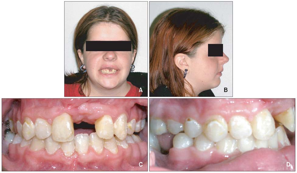
Figure 8 Final record of patient 2 at age 18 years. A, Frontal facial view; B, lateral facial view; C, frontal intraoral view; D, lateral intraoral view.
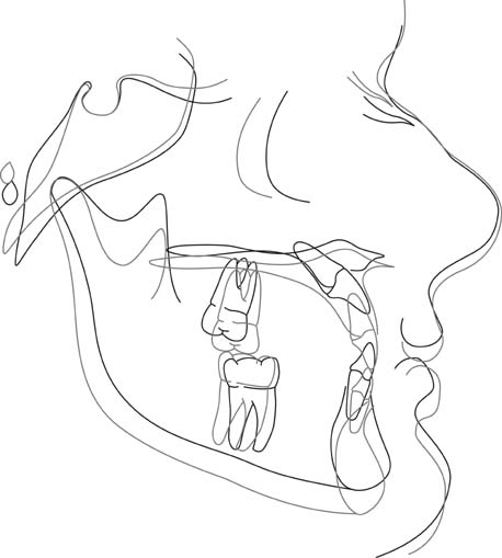
Figure 9 Superimposition of the pretreatment (dotted line) and posttreatment (solid line) lateral cephalograms in case 2.
Cited by 1 articles
-
Complex dental anomalies in a belatedly diagnosed cleidocranial dysplasia patient
Hui Lu, Binghui Zeng, Dongsheng Yu, Xiangyi Jing, Bin Hu, Wei Zhao, Yiming Wang
Imaging Sci Dent. 2015;45(3):187-192. doi: 10.5624/isd.2015.45.3.187.
Reference
-
1. Ishii K, Nielsen IL, Vargervik K. Characteristics of jaw growth in cleidocranial dysplasia. Cleft Palate Craniofac J. 1998; 35:161–166.
Article2. Lou Y, Javed A, Hussain S, Colby J, Frederick D, Pratap J, et al. A Runx2 threshold for the cleidocranial dysplasia phenotype. Hum Mol Genet. 2009; 18:556–568.
Article3. Mundlos S, Otto F, Mundlos C, Mulliken JB, Aylsworth AS, Albright S, et al. Mutations involving the transcription factor CBFA1 cause cleidocranial dysplasia. Cell. 1997; 89:773–779.
Article4. Ducy P, Zhang R, Geoffroy V, Ridall AL, Karsenty G. Osf2/Cbfa1: a transcriptional activator of osteoblast differentiation. Cell. 1997; 89:747–754.
Article5. Baumert U, Golan I, Redlich M, Aknin JJ, Muessig D. Cleidocranial dysplasia: molecular genetic analysis and phenotypic-based description of a Middle European patient group. Am J Med Genet A. 2005; 139A:78–85.
Article6. Yamashiro T, Aberg T, Levanon D, Groner Y, Thesleff I. Expression of Runx1, -2 and -3 during tooth, palate and craniofacial bone development. Mech Dev. 2002; 119:Suppl 1. S107–S110.
Article7. Lossdörfer S, Yildiz F, Götz W, Kheralla Y, Jäger A. Anabolic effect of intermittent PTH(1-34) on the local microenvironment during the late phase of periodontal repair in a rat model of tooth root resorption. Clin Oral Investig. 2010; 14:89–98.
Article8. Manjunath K, Kavitha B, Saraswathi TR, Sivapathasundharam B, Manikandhan R. Cementum analysis in cleidocranial dysostosis. Indian J Dent Res. 2008; 19:253–256.
Article9. Counts AL, Rohrer MD, Prasad H, Bolen P. An assessment of root cementum in cleidocranial dysplasia. Angle Orthod. 2001; 71:293–298.10. Tanaka JL, Ono E, Filho EM, Castilho JC, Moraes LC, Moraes ME. Cleidocranial dysplasia: importance of radiographic images in diagnosis of the condition. J Oral Sci. 2006; 48:161–166.
Article11. Suda N, Hattori M, Kosaki K, Banshodani A, Kozai K, Tanimoto K, et al. Correlation between genotype and supernumerary tooth formation in cleidocranial dysplasia. Orthod Craniofac Res. 2010; 13:197–202.
Article12. Becker A, Lustmann J, Shteyer A. Cleidocranial dysplasia: Part 1-General principles of the orthodontic and surgical treatment modality. Am J Orthod Dentofacial Orthop. 1997; 111:28–33.
Article13. Roberts T, Stephen L, Beighton P. Cleidocranial dysplasia: a review of the dental, historical, and practical implications with an overview of the South African experience. Oral Surg Oral Med Oral Pathol Oral Radiol. 2013; 115:46–55.
Article14. Liu DG, Zhang WL, Zhang ZY, Wu YT, Ma XC. Three-dimensional evaluations of supernumerary teeth using cone-beam computed tomography for 487 cases. Oral Surg Oral Med Oral Pathol Oral Radiol Endod. 2007; 103:403–411.
Article
