Korean J Orthod.
2013 Oct;43(5):225-234. 10.4041/kjod.2013.43.5.225.
Evaluation of the relationship between upper incisor exposure and cephalometric variables in Korean young adults
- Affiliations
-
- 1Department of Orthodontics, College of Dentistry, Wonkwang University, Iksan, Korea. pigtail@wku.ac.kr
- 2Department of Orthodontics, Wonkwang University Sanbon Dental Hospital, College of Dentistry, Wonkwang University, Gunpo, Korea.
- 3Department of Orthodontics, Wonkwang University Daejeon Dental Hospital, College of Dentistry, Wonkwang University, Daejeon, Korea.
- KMID: 2273414
- DOI: http://doi.org/10.4041/kjod.2013.43.5.225
Abstract
OBJECTIVE
The purpose of this study was to classify Korean young adults into 3 groups on the basis of upper incisor exposure rates (UIERs) and to compare the skeletal, dental, and soft tissue variables.
METHODS
Samples were obtained from 127 students at the College of Dentistry, Wonkwang University in South Korea. Facial photographs of frontal posed smiles and lateral cephalograms of the subjects were taken. The subjects were divided into 3 groups on the basis of UIERs and 20 measurements were compared among the 3 groups. The correlations between the variables were determined.
RESULTS
Male and female subjects showed significant differences in the group distribution. Male subjects showed higher frequencies of low smiles, and female subjects showed higher frequencies of high smiles. The vertical height of the anterior alveolar process of the maxilla directly correlated with the UIER. However, the UIER showed no significant correlation with the vertical height of the anterior basal bone or the inclination of the upper incisor axis. In female subjects, the upper central incisor clinical crown length showed an inverse correlation with the UIER. However, this variable showed no significant correlation with the UIER in male subjects.
CONCLUSIONS
The UIER was directly correlated with the levator muscle activity of the upper lip and inversely correlated with the upper lip thickness, yet there was no correlation between the UIER and upper lip length at rest.
Keyword
MeSH Terms
Figure
-
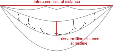
Figure 1 Modified smile index = (intervermillion distance at midline / intercommissural distance) ×100.

Figure 2 Facial index: N'-Gn'/ Zy'-Zy'; Upper facial index: N'-Sto'/ Zy'-Zy'. See Table 1 for the definitions of all the landmarks and measurements.
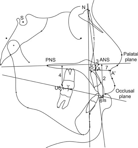
Figure 3 Cephalometric linear measurements. 1, Vertical-A; 2, PP-Is; 3, PP-A; 4, PP-U6; 5, A-N perpend; 6, NA-Is; 7, upper lip thickness. See Table 1 for the definitions of all the landmarks and measurements.
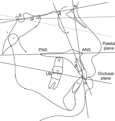
Figure 4 Cephalometric angular measurements. 1, SNA; 2, SN-OP; 3, SN-PP; 4, SN-U1; 5, NA-U1. Dashed and dotted lines indicate parallel planes to the palatal and occlusal planes, respectively. See Table 1 for the definitions of all the landmarks and measurements.
Reference
-
1. Tjan AH, Miller GD, The JG. Some esthetic factors in a smile. J Prosthet Dent. 1984; 51:24–28.
Article2. LaFrance M, Hecht MA, Paluck EL. The contingent smile: a meta-analysis of sex differences in smiling. Psychol Bull. 2003; 129:305–334.
Article3. Sarver DM. The importance of incisor positioning in the esthetic smile: the smile arc. Am J Orthod Dentofacial Orthop. 2001; 120:98–111.
Article4. Rigsbee OH 3rd, Sperry TP, BeGole EA. The influence of facial animation on smile characteristics. Int J Adult Orthodon Orthognath Surg. 1988; 3:233–239.5. Sarver DM, Ackerman MB. Dynamic smile visualization and quantification: Part 2. Smile analysis and treatment strategies. Am J Orthod Dentofacial Orthop. 2003; 124:116–127.
Article6. Ackerman JL, Ackerman MB, Brensinger CM, Landis JR. A morphometric analysis of the posed smile. Clin Orthod Res. 1998; 1:2–11.
Article7. Hulsey CM. An esthetic evaluation of lip-teeth relationships present in the smile. Am J Orthod. 1970; 57:132–144.
Article8. Ackerman MB, Ackerman JL. Smile analysis and design in the digital era. J Clin Orthod. 2002; 36:221–236.9. Proffit WR, Fields HW, Sarver DM. Contemporary orthodontics. 4th ed. St. Louis: Mosby Elsevier;2007.10. Schabel BJ, Baccetti T, Franchi L, McNamara JA. Clinical photography vs digital video clips for the assessment of smile esthetics. Angle Orthod. 2010; 80:490–496.
Article11. Dong JK, Jin TH, Cho HW, Oh SC. The esthetics of the smile: a review of some recent studies. Int J Prosthodont. 1999; 12:9–19.12. Peck S, Peck L, Kataja M. The gingival smile line. Angle Orthod. 1992; 62:91–100.13. Krishnan V, Daniel ST, Lazar D, Asok A. Characterization of posed smile by using visual analog scale, smile arc, buccal corridor measures, and modified smile index. Am J Orthod Dentofacial Orthop. 2008; 133:515–523.
Article14. Peck S, Peck L, Kataja M. Some vertical lineaments of lip position. Am J Orthod Dentofacial Orthop. 1992; 101:519–524.
Article15. Vig RG, Brundo GC. The kinetics of anterior tooth display. J Prosthet Dent. 1978; 39:502–504.
Article16. Farkas LG, Posnick JC, Hreczko TM. Growth patterns of the face: a morphometric study. Cleft Palate Craniofac J. 1992; 29:308–315.
Article17. Dahlberg S. Statistical methods for medical and biological students. New York: Interscience;1940.18. Sarver DM, Ackerman MB. Dynamic smile visualization and quantification: part 1. Evolution of the concept and dynamic records for smile capture. Am J Orthod Dentofacial Orthop. 2003; 124:4–12.
Article19. Shafiee R, Korn EL, Pearson H, Boyd RL, Baumrind S. Evaluation of facial attractiveness from end-of-treatment facial photographs. Am J Orthod Dentofacial Orthop. 2008; 133:500–508.
Article20. Mamandras AH. Linear changes of the maxillary and mandibular lips. Am J Orthod Dentofacial Orthop. 1988; 94:405–410.
Article
- Full Text Links
-
- Actions
-
Cited
- CITED
-
- Close
- Share
-
- Similar articles
-
- A study of upper and lower incisor position in normal occlusion
- A study on the affecting factors on root resorption
- The Esthetic Upper Incisor Position in Korean Adult Female
- A cephalometric study of soft tissue profile changes associated with orthodontic treatment
- Evaluation of the lip during smile in normal occlusion adults
