Distortion of tooth axes on panoramic radiographs taken at various head positions
- Affiliations
-
- 1Department of Orthodontics, College of Dentistry, Chosun University, Korea. shlim@chosun.ac.kr
- KMID: 2273365
- DOI: http://doi.org/10.4041/kjod.2008.38.4.240
Abstract
OBJECTIVE
The purpose of this study was to evaluate the effect of head position changes on the root parallelism between adjacent teeth on panoramic radiographs. METHODS: A model with normal occlusion was constructed in the SolidWorks program, then RP (rapid protyping) model was fabricated. The model was repeatedly imaged and repositioned five times at each of the following nine positions: ideal head position, 5degrees C up, 10degrees C up, 5degrees C down, 10degrees C down, 5degrees C right, 10degrees C right up, and 5degrees C right rotation, 10degrees C right rotation. Panoramic radiographs were taken by Planmeca ProMax and the angle between the long axes of adjacent teeth was directly measured in the monitor. RESULTS: Axes of adjacent teeth tended to converge toward the occlusal plane when the head tilted up and converged in the opposite direction to the occlusal plane when the head tilted down. Anterior teeth showed the most notable differences. When one side of the head tilted up 5degrees C and 10degrees C along the anteroposterior axis (Y axis), tooth axes of the same side tended to converge toward the occlusal plane and tooth axes of the opposite side tended to converge in the opposite direction to the occlusal plane. When the head rotated to one side along the vertical axis (Z axis), the canine and lateral incisor of the same side converged in the opposite direction to the occlusal plane and the canine and lateral incisor of the other side converged toward the occlusal plane. CONCLUSIONS: When assessing the root parallelism on panoramic radiographs, the occlusal plane cant (anteroposterior or lateral) or asymmetry of the dental arch should be considered because these can cause distortion of tooth axes on panoramic radiographs.
MeSH Terms
Figure
-
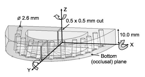
Fig 1 3D model and axes of rotation that were used in this study. X, transverse axis; Y, anteroposterior axis; Z, vertical axis. The model was constructed using SolidWorks program. Arrows show the model being tilted anteroposteriorly along the transverse axis (X axis), tilted laterally along the anteroposterior axis (Y axis), and rotated laterally along the vertical axis (Z axis).
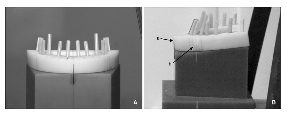
Fig 2 Model positioning in the panoramic radiograph machine. A, Frontal view; B, lateral view; midsagittal positioning beam (a) was aligned to model's midline and focal layer positioning beam (b) was aligned between the upper lateral incisor and upper canine.

Fig 3 Angular measurement of panoramic radiographs using the measurement tool (Cobb's angle) of the π-ViewStar program. a, Angle between long axes of adjacent teeth was measured on the occlusal side when long axes of adjacent teeth was converged toward the occlusal plane; b, angle between long axes of adjacent teeth was measured on the apical side when long axes of adjacent teeth were converged in the opposite direction to the occlusal plane. This was assigned a negative value (??.

Fig 4 Angle between long axes of adjacent teeth at ideal head position and head positions tilted anteroposteriorly along the transverse axis (5° up, 10° up, 5° down, and 10° down). Values with (?? indicate that the long axes of adjacent teeth were converged in the opposite direction to the occlusal plane.
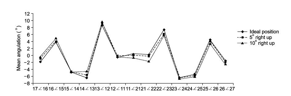
Fig 5 Angle between long axes of adjacent teeth at ideal head position and head positions tilted laterally along the anteroposterior axis (5° right up and 10° right up). Values with (-) indicate that the long axes of adjacent teeth were converged in the opposite direction to the occlusal plane.
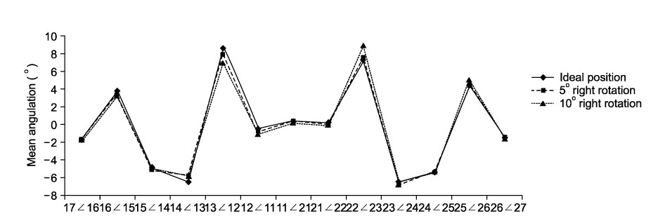
Fig 6 Angle between long axes of adjacent teeth at ideal head position and head positions rotated laterally along the vertical axis (5° right rotation and 10° right rotation). Values with (-) indicate that the long axes of adjacent teeth were converged in the opposite direction to the occlusal plane.
Cited by 1 articles
-
Evaluation of mesiodistal tooth axis using a CBCT-generated panoramic view
In-Tae Song, Jin-Hyoung Cho, Jong-Moon Chae, Na-Young Chang
Korean J Orthod. 2011;41(4):255-267. doi: 10.4041/kjod.2011.41.4.255.
Reference
-
1. Frykholm A, Malmgren O, Sämfors KA, Welander U. Angular measurements in orthopantomography. Dentomaxillofac Radiol. 1977. 6:77–81.
Article2. Floyd P, Palmer P, Palmer R. Radiographic techniques. Br Dent J. 1999. 187:359–365.
Article3. Wyatt CC, Pharoah MJ. Imaging techniques and image interpretation for dental implant treatment. Int J Prosthodont. 1998. 11:442–452.4. Lam EW, Ruprecht A, Yang J. Comparison of two-dimensional orthoradially reformatted computed tomography and panoramic radiography for dental implant treatment planning. J Prosthet Dent. 1995. 74:42–46.
Article5. Frederiksen NL. Diagnostic imaging in dental implantology. Oral Surg Oral Med Oral Pathol Oral Radiol Endod. 1995. 80:540–554.
Article6. Holdaway RA. Bracket angulation as applied to the edgewise appliance. Angle Orthod. 1952. 22:227–236.7. Mayoral G. Treatment results with light wires studied by panoramic radiography. Am J Orthod. 1982. 81:489–497.
Article8. Jarabak JR, Fizzell JA. Technique and treatment with lightwire edgewise appliances. 1972. St Louis: Mosby;277–379.9. Hatasaka HH. A radiographic study of roots in extraction sites. Angle Orthod. 1976. 46:64–68.10. Strang RJ. Factors associated with successful orthodontic treatment. Am J Orthod. 1952. 38:790–800.
Article11. Lucchesi MV, Wood RE, Nortjé CJ. Suitability of the panoramic radiograph for assessment of mesiodistal angulation of teeth in the buccal segments of the mandible. Am J Orthod Dentofacial Orthop. 1988. 94:303–310.
Article12. Casko JS, Vaden JL, Kokich VG, Damone J, James RD, Cangialosi TJ, et al. Objective grading system for dental casts and panoramic radiographs. American Board of Orthodontics. Am J Orthod Dentofacial Orthop. 1998. 114:589–599.13. Ursi WJ, Almeida RR, Tavano O, Henriques JF. Assessment of mesiodistal axial inclination through panoramic radiography. J Clin Orthod. 1990. 24:166–173.14. Xie Q, Soikkonen K, Wolf J, Mattila K, Gong M, Ainamo A. Effect of head positioning in panoramic radiography on vertical measurements: an in vitro study. Dentomaxillofac Radiol. 1996. 25:61–66.
Article15. Sämfors KA, Welander U. Angle distortion in narrow beam rotation radiography. Acta Radiol Diagn (Stockh). 1974. 15:570–576.
Article16. Tronje G, Welander U, McDavid WD, Morris CR. Image distortion in rotational panoramic radiography. III. Inclined objects. Acta Radiol Diagn Stockh. 1981. 22:585–592.17. McDavid WD, Tronje G, Welander U, Morris CR, Nummikoski P. Imaging characteristics of seven panoramic x-ray units. Dentomaxillofac Radiol. 1985. 8:(suppl). 29S–34S.18. Larheim TA, Svanaes DB, Johannessen S. Reproducibility of radiographs with the Orthopantomograph 5: tooth-length assessment. Oral Surg Oral Med Oral Pathol. 1984. 58:736–741.
Article19. Larheim TA, Svanaes DB. Reproducibility of rotational panoramic radiography: mandibular linear dimensions and angles. Am J Orthod Dentofacial Orthop. 1986. 90:45–51.
Article20. Mckee LW, Glover KE, Williamson PC, Lam EW, Heo G, Major PW. The effect of vertical and horizontal head positioning in panoramic radiography on mesiodistal tooth angulations. Angle Orthod. 2001. 71:442–451.21. Stramotas S, Geenty JP, Petocz P, Darendeliler MA. Accuracy of linear and angular measurements on panoramic radiographs taken at various positions in vitro. Eur J Orthod. 2002. 24:43–52.
Article22. Schiff T, D'Ambrosio J, Glass BJ, Langlais RP, McDavid WD. Common positioning and technical errors in panoramic radiography. J Am Dent Assoc. 1986. 113:422–426.
Article23. Choi GL. Accuracy of mesiodistal angulation of teeth in panoramic radiographs [thesis]. 2005. Gwangju: Chosun University.24. Ash MM. Wheeler's dental anatomy physiology, and occlusion. 1984. Pennsylvania: Saunders Company;387–392.25. Cohen S, Burns RC. Pathways of the pulp. 2003. St Louis: Mosby;151.26. Ricketts RM. Cephalometric analysis synthesis. Angle Orthod. 1961. 31:141–156.27. Ramstad T, Hensten-Pettersen O, Mohn E, Ibrahim SI. A methodological study of errors in vertical measurements of edentulous ridge height on orthopantomographic radiograms. J Oral Rehabil. 1978. 5:403–412.
Article28. Türp JC, Vach W, Harbich K, Alt KW, Strub JR. Determiningmandibular condyle and ramus height with the help of an orthopantomogram- a valid method? J Oral Rehabil. 1996. 23:395–400.29. Batenburg RH, Stellingsma K, Raghoebar GM, Vissink A. Bone height measurements on panoramic radiographs: the effect of shape and position of edentulous mandibles. Oral Surg Oral Med Oral Pathol Oral Radiol Endod. 1997. 84:430–435.30. Kim JD, Kim JS, You CH. Mesiodistal tooth angulation to segmental occlusal plane in panoramic radiography. Korean J Oral Maxillofac Radiol. 2005. 35:25–31.31. Philipp RG, Hurst RV. The cant of the occlusal plane and distortion in the panoramic radiograph. Angle Orthod. 1978. 48:317–323.32. Samawi SS, Burke PH. Angular distortion in the orthopantomogram. Br J Orthod. 1984. 11:100–107.
Article33. Yoon YJ, Kim DH, Yu PS, Kim HJ, Choi EH, Kim KW. Effect of head rotation on posteroanterior cephalometric radiographs. Angle Orthod. 2002. 72:36–42.
- Full Text Links
-
- Actions
-
Cited
- CITED
-
- Close
- Share
-
- Similar articles
-
- Mesiodistal tooth angulation to segmental occlusal plane in panoramic radiography
- Evaluation of mesiodistal tooth axis using a CBCT-generated panoramic view
- Evaluation of the accuracy of linear and angular measurements on panoramic radiographs taken at different positions
- Quantitative localization of impacted mesiodens using panoramic and periapical radiographs
- The radiographic localization of unerupted maxillary incisors and supernumeraries
