Interdisciplinary rehabilitation of a root-fractured maxillary central incisor: A 12-year follow-up case report
- Affiliations
-
- 1Unit of Orthodontics, Department of Biomedical and Neuromotor Sciences (DIBINEM), University of Bologna, Italy. giulio.alessandri@unibo.it
- 2Private Practice, Faenza (Ravenna), Italy.
- 3Unit of Periodontology, DIBINEM, University of Bologna, Italy.
- KMID: 2273278
- DOI: http://doi.org/10.4041/kjod.2014.44.4.217
Abstract
- Single-tooth implantation has become a common treatment solution for replacement of a root-fractured maxillary incisor in adults, but the long-term esthetic results can be unfavorable due to progressive marginal bone loss, resulting in gingival recession. In this case report, a maxillary central incisor with a root fracture in its apical one-third was orthodontically extruded and extracted in a 21-year-old female. Implant surgery was performed after a 3-month healing period, and the final crown was placed about 12 months after extraction. After 12 years, favorable osseous and gingival architectures were visible with adequate bone height and thickness at the buccal cortical plate, and no gingival recession was seen around the implant-supported crown. Although modern dentistry has been shifting toward simplified, clinical procedures and shorter treatment times, both general dentists and orthodontists should be aware of the possible long-term esthetic advantages of orthodontic extrusion of hopelessly fractured teeth for highly esthetically demanding areas and should educate and motivate patients regarding the choice of this treatment solution, if necessary.
MeSH Terms
Figure
-
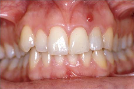
Figure 1 Pretreatment intraoral photograph.

Figure 2 Pretreatment periapical radiograph. Note the horizontal root fracture of the left central incisor and the circular radiolucency around the apex of the adjacent lateral incisor.
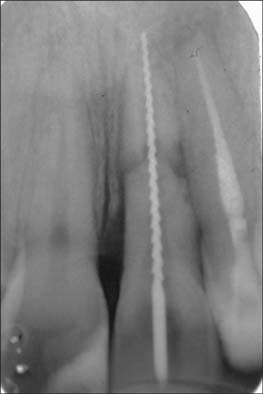
Figure 3 A periapical radiograph that was obtained after the lateral incisor had endodontic treatment. Moreover, note that a hand file was cemented into the root canal of the central incisor before orthodontic extrusion.
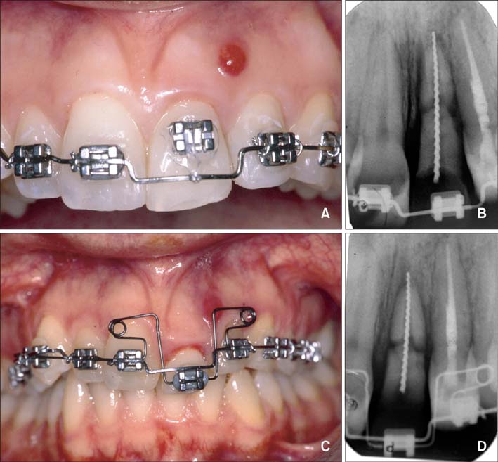
Figure 4 Orthodontic extrusion of the root-fractured central incisor: clinical (A) and radiographic (B) views after bracket placement and elastic chain application; clinical (C) and radiographic (D) views after adoption of a multiple-loop design orthodontic appliance.

Figure 5 After a 4-month orthodontic extrusion, followed by a 3-month retention phase: clinical (A) and radiographic (B) views after appliance removal.

Figure 6 Clinical (A) and radiographic (B) views after tooth extraction and cementation of a Maryland bridge on the teeth adjacent to the extraction site.

Figure 7 Intraoral frontal view (A) and periapical radiograph (B) obtained after placement of implant and its final metal-ceramic crown.

Figure 8 Intraoral frontal view (A) and periapical radiograph (B) obtained at 6-year follow-up.
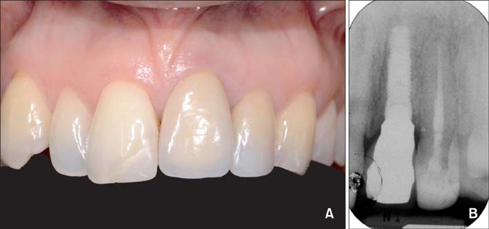
Figure 9 Intraoral frontal view (A) and periapical radiograph (B) obtained at 12-year follow-up.

Figure 10 Cone-beam computed tomography images were obtained at the 12-year follow-up: axial (A) and sagittal (B) views show the presence of a well-represented buccal cortical plate.
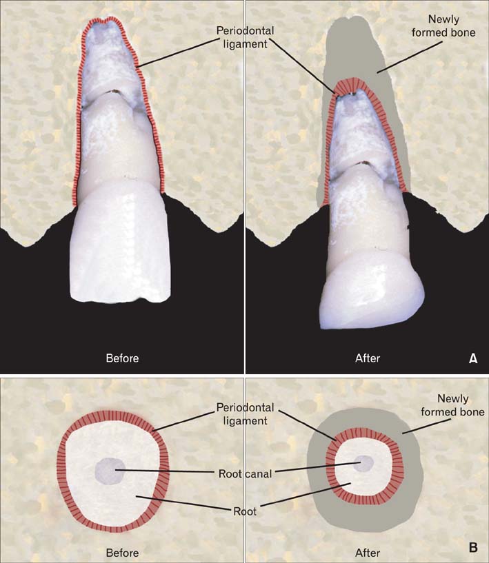
Figure 11 Graphical representation of the cross section of the root-fractured maxillary central incisor before and after orthodontic extrusion: (A) frontal view; (B) axial view at the level of the cementoenamel junction of the adjacent central incisor.
Reference
-
1. Buser D, Chen ST, Weber HP, Belser UC. Early implant placement following single-tooth extraction in the esthetic zone: biologic rationale and surgical procedures. Int J Periodontics Restorative Dent. 2008; 28:441–451.2. Mijiritsky E, Mardinger O, Mazor Z, Chaushu G. Immediate provisionalization of single-tooth implants in fresh-extraction sites at the maxillary esthetic zone: up to 6 years of follow-up. Implant Dent. 2009; 18:326–333.
Article3. Buser D, Halbritter S, Hart C, Bornstein MM, Grütter L, Chappuis V, et al. Early implant placement with simultaneous guided bone regeneration following single-tooth extraction in the esthetic zone: 12-month results of a prospective study with 20 consecutive patients. J Periodontol. 2009; 80:152–162.
Article4. Gozneli R, Ozkan Y, Akalin ZF, Ozkan Y. Rehabilitation of maxillary anterior esthetics by alveolar distraction osteogenesis with immediate implant placement: a case report. Implant Dent. 2010; 19:468–476.
Article5. Zachrisson BU, Toreskog BU. Esthetic considerations in restoring the traumatized dentition: a biologic approach. In : Andreasen JO, Andreasen FM, Andersson L, editors. Textbook and color atlas of traumatic injuries to the teeth. Copenhagen: Blackwell Munksgaard;2007. p. 798–813.6. de Molon RS, de Avila ED, de Souza JA, Nogueira AV, Cirelli CC, Margonar R, et al. Forced orthodontic eruption for augmentation of soft and hard tissue prior to implant placement. Contemp Clin Dent. 2013; 4:243–247.
Article7. Schropp L, Wenzel A, Kostopoulos L, Karring T. Bone healing and soft tissue contour changes following single-tooth extraction: a clinical and radiographic 12-month prospective study. Int J Periodontics Restorative Dent. 2003; 23:313–323.8. Cardaropoli G, Araújo M, Lindhe J. Dynamics of bone tissue formation in tooth extraction sites. An experimental study in dogs. J Clin Periodontol. 2003; 30:809–818.9. Maiorana C, Speroni S, Herford AS, Cicciù M. Slow orthodontic teeth extrusion to enhance hard and soft periodontal tissue quality before implant positioning in aesthetic area. Open Dent J. 2012; 6:137–142.
Article10. Reitan K. Clinical and histologic observations on tooth movement during and after orthodontic treatment. Am J Orthod. 1967; 53:721–745.
Article11. Salama H, Salama M. The role of orthodontic extrusive remodeling in the enhancement of soft and hard tissue profiles prior to implant placement: a systematic approach to the management of extraction site defects. Int J Periodontics Restorative Dent. 1993; 13:312–333.12. Bahadure RN, Thosar N, Khubchandani M. Orthodontic extrusion: diagnosis and treatment with CBCT in a pediatric patient. Gen Dent. 2013; 61:e5–e7.13. Chambrone L, Chambrone LA. Forced orthodontic eruption of fractured teeth before implant placement: case report. J Can Dent Assoc. 2005; 71:257–261.14. Amato F, Mirabella AD, Macca U, Tarnow DP. Implant site development by orthodontic forced extraction: a preliminary study. Int J Oral Maxillofac Implants. 2012; 27:411–420.15. Kan JY, Rungcharassaeng K, Umezu K, Kois JC. Dimensions of peri-implant mucosa: an evaluation of maxillary anterior single implants in humans. J Periodontol. 2003; 74:557–562.
Article16. Mantzikos T, Shamus I. Forced eruption and implant site development: soft tissue response. Am J Orthod Dentofacial Orthop. 1997; 112:596–606.
Article17. Weisgold AS, Arnoux JP, Lu J. Single-tooth anterior implant: a world of caution. Part I. J Esthet Dent. 1997; 9:225–233.18. Benic GI, Mokti M, Chen CJ, Weber HP, Hämmerle CH, Gallucci GO. Dimensions of buccal bone and mucosa at immediately placed implants after 7 years: a clinical and cone beam computed tomography study. Clin Oral Implants Res. 2012; 23:560–566.
Article19. Chang M, Wennström JL, Odman P, Andersson B. Implant supported single-tooth replacements compared to contralateral natural teeth. Crown and soft tissue dimensions. Clin Oral Implants Res. 1999; 10:185–194.
Article20. Esposito M, Grusovin MG, Polyzos IP, Felice P, Worthington HV. Interventions for replacing missing teeth: dental implants in fresh extraction sockets (immediate, immediate-delayed and delayed implants). Cochrane Database Syst Rev. 2010; (9):CD005968.
Article21. Rominger JW, Triplett RG. The use of guided tissue regeneration to improve implant osseointegration. J Oral Maxillofac Surg. 1994; 52:106–112.
Article22. Bonetti GA, Parenti SI, Checchi L. Orthodontic extraction of mandibular third molar to avoid nerve injury and promote periodontal healing. J Clin Periodontol. 2008; 35:719–723.
Article23. Zucchelli G, Parenti SI, Ghigi G, Bonetti GA. Combined orthodontic - mucogingival treatment of a deep post-orthodontic gingival recession. Eur J Esthet Dent. 2012; 7:266–280.24. Alessandri Bonetti G, Incerti Parenti S, Zucchelli G. Onychophagia and postorthodontic isolated gingival recession: diagnosis and treatment. Am J Orthod Dentofacial Orthop. 2012; 142:872–878.
Article25. Belser UC, Grütter L, Vailati F, Bornstein MM, Weber HP, Buser D. Outcome evaluation of early placed maxillary anterior single-tooth implants using objective esthetic criteria: a cross-sectional, retrospective study in 45 patients with a 2- to 4-year follow-up using pink and white esthetic scores. J Periodontol. 2009; 80:140–151.
Article26. Buser D, Wittneben J, Bornstein MM, Grütter L, Chappuis V, Belser UC. Stability of contour augmentation and esthetic outcomes of implant-supported single crowns in the esthetic zone: 3-year results of a prospective study with early implant placement postextraction. J Periodontol. 2011; 82:342–349.
Article
- Full Text Links
-
- Actions
-
Cited
- CITED
-
- Close
- Share
-
- Similar articles
-
- Use of mineral trioxide aggregate in the treatment of horizontal root fracture with a 4-year follow-up: case report
- Treatment of horizontal root-fractured maxillary incisors
- Orthodontic treatment of an impacted maxillary central incisor with dilacerations
- Unilateral maxillary central incisor root resorption after orthodontic treatment for Angle Class II, division 1 malocclusion with significant maxillary midline deviation: A possible correlation with root proximity to the incisive canal
- Healing after horizontal root fractures: 3 cases with 2-year follow-up
