The effect of bonded resin surface area on the detachment force of lingual bonded fixed retainers: An in vitro study
- Affiliations
-
- 1Department of Orthodontics, Hallym University Sacred Heart Hospital, Hallym University Medical Center, Anyang, Korea.
- 2Department and Research Institute of Dental Biomaterials and Bioengineering, College of Dentistry, Yonsei University, Seoul, Korea.
- 3Department of Orthodontics, Hallym University Kangnam Sacred Heart Hospital, Hallym University Medical Center, Seoul, Korea. ajh0225@gmail.com
- KMID: 2273267
- DOI: http://doi.org/10.4041/kjod.2014.44.1.20
Abstract
OBJECTIVE
The aims of this study were to evaluate the relationship between the detachment force and bonding resin surface are and to determine the resin bonding surface area that would provide adequate bonding strength with minimum resin volume.
METHODS
One hundred and sixty human premolars were randomly divided into 4 groups of 40 teeth each. The diameter of the resin surface area in each group was as follows: group 1, 1.5 mm; group 2, 2.5 mm; group 3, 3.5 mm; and group 4, 4.5 mm. Respond Dead Soft straight (length 0.0175 inch) was used to fabricate the retainers, and Transbond(TM) XT was used to fix the retainers to the tooth surfaces. A pair of teeth was embedded in acrylic blocks for each specimen. Thus, each group comprised 20 samples. Fixed retainers were bonded to the teeth, and vertical force was applied at the middle of wire. The force was measured using a universal testing machine.
RESULTS
The mean value of detachment force was the highest for group 4 (102.38 +/- 2.92 N), followed by group 3 (63.54 +/- 2.21 N), group 2 (51.95 +/- 1.61 N), and group 1 (24.14 +/- 1.38 N).
CONCLUSIONS
The detachment force of lingual fixed retainers was significantly affected as the area of the resin bonding surface increased. Considering the minimum bonding strength of brackets, a resin bonding surface area with a diameter of 3.5 mm would provide adequate bonding strength.
Figure
-
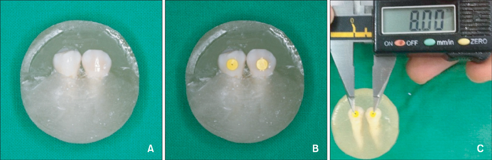
Figure 1 The experimental sample. A, One pair of teeth embedded in a resin block. B, A sticker attached to the buccal surfaces of the sample teeth. C, The distance between the midpoints of 2 stickers (8 mm).
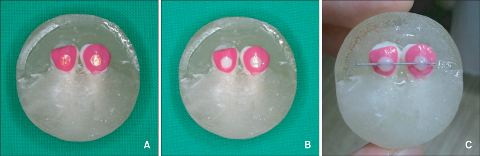
Figure 2 The preparation of the resin bonding surface. A, The surfaces of the teeth were covered by stickers and surrounded by nail polish. B, The resin bonding surface after removal of the sticker. C, A wire is attached to the tooth surfaces with bonding resin.

Figure 3 The universal testing machine. The chisel-edge plunger was aimed at the center of the wire and was parallel to the long axis of the teeth.
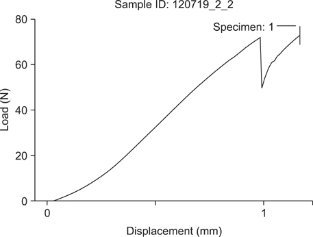
Figure 4 The relationship between the force applied to the wire and displacement of the wire. The detachment force was measured when the force perimeter dropped rapidly.

Figure 5 Means and standard deviations of the detachment forces for each group. Group 1, resin surface diameter = 1.5 mm; Group 2, 2.5 mm; Group 3, 3.5 mm; Group 4, 4.5 mm.
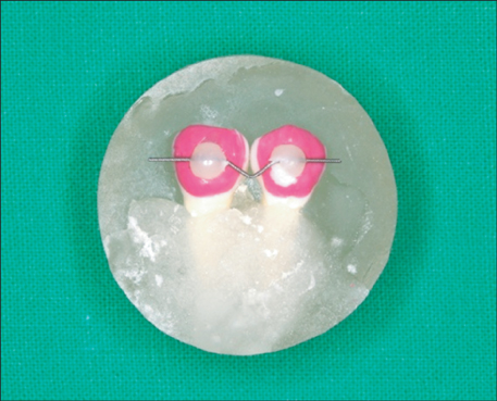
Figure 6 A sample showing a "V"-shaped deformation after detachment.
Reference
-
1. Little RM, Riedel RA, Artun J. An evaluation of changes in mandibular anterior alignment from 10 to 20 years postretention. Am J Orthod Dentofacial Orthop. 1988; 93:423–428.
Article2. Little RM, Riedel RA, Engst ED. Serial extraction of first premolars-postretention evaluation of stability and relapse. Angle Orthod. 1990; 60:255–262.3. Little RM, Wallen TR, Riedel RA. Stability and relapse of mandibular anterior alignment-first premolar extraction cases treated by traditional edgewise orthodontics. Am J Orthod. 1981; 80:349–365.
Article4. Littlewood SJ, Millett DT, Doubleday B, Bearn DR, Worthington HV. Retention procedures for stabilising tooth position after treatment with orthodontic braces. Cochrane Database Syst Rev. 2006; (1):CD002283.
Article5. Nanda RS, Nanda SK. Considerations of dentofacial growth in long-term retention and stability: is active retention needed? Am J Orthod Dentofacial Orthop. 1992; 101:297–302.
Article6. Zachrisson BU. Clinical experience with direct-bonded orthodontic retainers. Am J Orthod. 1977; 71:440–448.
Article7. Zachrisson BU. Adults retention, a new approach in orthdontics. In : Graber LW, Graber TM, editors. Orthodontics, state of the art, essence of the science. St. Louis: Mosby;1986.8. Bearn DR, McCabe JF, Gordon PH, Aird JC. Bonded orthodontic retainers: the wire-composite interface. Am J Orthod Dentofacial Orthop. 1997; 111:67–74.
Article9. Cooke ME, Sherriff M. Debonding force and deformation of two multi-stranded lingual retainer wires bonded to incisor enamel: an in vitro study. Eur J Orthod. 2010; 32:741–746.
Article10. Baysal A, Uysal T, Gul N, Alan MB, Ramoglu SI. Comparison of three different orthodontic wires for bonded lingual retainer fabrication. Korean J Orthod. 2012; 42:39–46.
Article11. Kang DK, Son WS, Kim HI. Bond strength of bonded lingual retainer with flowable composite resin. J Korea Res Soc Dent Mater. 2004; 31:283–290.12. Artun J, Bergland S. Clinical trials with crystal growth conditioning as an alternative to acid-etch enamel pretreatment. Am J Orthod. 1984; 85:333–340.
Article13. Al Yami EA, Kuijpers-Jagtman AM, van't Hof MA. Stability of orthodontic treatment outcome: follow-up until 10 years postretention. Am J Orthod Dentofacial Orthop. 1999; 115:300–304.
Article14. Kwon TY, Meina H, Antoszewska J, Park HS. Direct and indirect bonding of wire retainers to bovine enamel using three resin systems: shear bond strength comparisons. Korean J Orthod. 2011; 41:447–453.
Article15. Oesterle LJ, Shellhart WC, Henderson S. Enhancing wire-composite bond strength of bonded retainers with wire surface treatment. Am J Orthod Dentofacial Orthop. 2001; 119:625–631.
Article16. Radlanski RJ, Zain ND. Stability of the bonded lingual wire retainer-a study of the initial bond strength. J Orofac Orthop. 2004; 65:321–335.
Article17. Dahlberg G. Statistical methods for medical and biological students. London: George Allen & Unwin;1940.18. Lee HC, Son WS. A study of shear bond strength of bonded retainer according to the bonding method and type of wires. Korean J Orthod. 2002; 32:143–153.19. Scougall Vilchis RJ, Yamamoto S, Kitai N, Yamamoto K. Shear bond strength of orthodontic brackets bonded with different self-etching adhesives. Am J Orthod Dentofacial Orthop. 2009; 136:425–430.
Article20. Hajrassie MK, Khier SE. In-vivo and in-vitro comparison of bond strengths of orthodontic brackets bonded to enamel and debonded at various times. Am J Orthod Dentofacial Orthop. 2007; 131:384–390.
Article21. Tavas MA, Watts DC. A visible light-activated direct bonding material: an in vitro comparative study. Br J Orthod. 1984; 11:33–37.
Article22. Murray SD, Hobson RS. Comparison of in vivo and in vitro shear bond strength. Am J Orthod Dentofacial Orthop. 2003; 123:2–9.
Article23. Bearn DR. Bonded orthodontic retainers: a review. Am J Orthod Dentofacial Orthop. 1995; 108:207–213.
Article24. Artun J, Urbye KS. The effect of orthodontic treatment on periodontal bone support in patients with advanced loss of marginal periodontium. Am J Orthod Dentofacial Orthop. 1988; 93:143–148.
Article
- Full Text Links
-
- Actions
-
Cited
- CITED
-
- Close
- Share
-
- Similar articles
-
- Clinical perspectives on 2-unit cantilevered resin-bonded fixed partial denture
- Effect of thermocycling on shear bond strength and mode of failure of bonded retainer using flowable composite resin
- The shear bond strength of two adhesives bonded to composite resin and glass ionomer cement restorations
- Comparison of three different orthodontic wires for bonded lingual retainer fabrication
- Resin-bonded fixed partial denture using In-Ceram and Targis-Ventris system
