Late development of a mandibular second premolar
- Affiliations
-
- 1Department of Orthodontics, Cumhuriyet University, Faculty of Dentistry, Sivas, Turkey. altugbicakci@gmail.com
- KMID: 2273262
- DOI: http://doi.org/10.4041/kjod.2012.42.2.94
Abstract
- In this report, we present the case of a girl with delayed odontogenesis of a lower second premolar for which she was followed up for 8.5 years. Congenital absence of permanent mandibular second premolars was observed at the initial radiographic examination at 8 years and 1 month. One year later, during the treatment period, an unexpected odontogenesis of a right second premolar was diagnosed on follow-up radiography. The original treatment plan was revised and a new plan was successfully implemented. Th is unusual case showed that the orthodontist's clinical philosophy must be flexible because unexpected situations can arise, especially when treating growing patients.
Figure
-
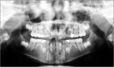
Figure 1 Panoramic radiograph of the patient aged 8 years and 1 month showing the absence of mandibular permanent second premolars.
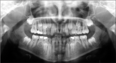
Figure 2 Panoramic radiograph of the patient aged 9 years and 6 months indicating mandibular right second premolar formation 17 months after initial diagnosis.
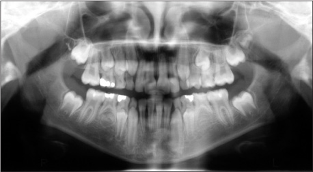
Figure 3 Panoramic radiograph of the patient showing no sign of mandibular left second premolar tooth formation, while calcification of the contralateral premolar had progressed.
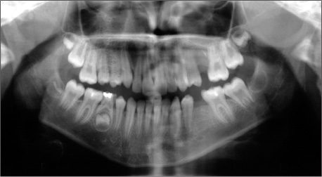
Figure 4 Panoramic radiograph of the patient demonstrating that mineralization of 3 third molars had begun while the forth one was in the germ phase; therefore, agenesis diagnosis for the left premolar was given and the deciduous molar was extracted.
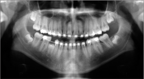
Figure 5 Panoramic radiograph of the patient showing that two-thirds of the late-developing premolar root formation was completed and the tooth was about to be exposed in the oral cavity.
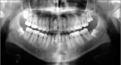
Figure 6 Panoramic radiograph of the patient aged 16 years and 6 months demonstrating that the mandibular right second premolar reached the occlusal plane at 8 years and 6 months after initial diagnosis. Note that the root formation was almost complete; however, the apex was still open.
Reference
-
1. Rølling S. Hypodontia of permanent teeth in Danish schoolchildren. Scand J Dent Res. 1980. 88:365–369.
Article2. Moyers RE. Handbook of orthodontics. 1972. 3rd ed. Chicago: Year Book Medical Publishers;166–239. 473–476.3. Fass EN. Aberrant second premolars. ASDC J Dent Child. 1970. 37:494–498.4. Moorrees CF, Fanning EA, Hunt EE Jr. Age variation of formation stages for ten permanent teeth. J Dent Res. 1963. 42:1490–1502.
Article5. Nolla CM. The development of the permanent teeth. J Dent Child. 1960. 27:254–266.6. Ravin JJ, Nielsen HG. A longitudinal radiographic study of the mineralization of 2nd premolars. Scand J Dent Res. 1977. 85:232–236.
Article7. Nielsen HG, Ravn JJ. A radiographic study of mineralization of permanent teeth in a group of children aged 3-7 years. Scand J Dent Res. 1976. 84:109–118.
Article8. Lindqvist B. Extraction of the deciduous second molar in hypodontia. Eur J Orthod. 1980. 2:173–181.
Article9. Nieminen P. Genetic basis of tooth agenesis. J Exp Zool B Mol Dev Evol. 2009. 312B:320–342.
Article10. Kist R, Watson M, Wang X, Cairns P, Miles C, Reid DJ, et al. Reduction of Pax9 gene dosage in an allelic series of mouse mutants causes hypodontia and oligodontia. Hum Mol Genet. 2005. 14:3605–3617.
Article11. Ranta R. Hypodontia and delayed development of the second premolars in cleft palate children. Eur J Orthod. 1983. 5:145–148.
Article12. Uslenghi S, Liversidge HM, Wong FS. A radiographic study of tooth development in hypodontia. Arch Oral Biol. 2006. 51:129–133.
Article13. Rune B, Sarnäs KV. Tooth size and tooth formation in children with advanced hypodontia. Angle Orthod. 1974. 44:316–321.14. Vastardis H, Karimbux N, Guthua SW, Seidman JG, Seidman CE. A human MSX1 homeodomain missense mutation causes selective tooth agenesis. Nat Genet. 1996. 13:417–421.
Article15. Stockton DW, Das P, Goldenberg M, D'Souza RN, Patel PI. Mutation of PAX9 is associated with oligodontia. Nat Genet. 2000. 24:18–19.
Article16. Garn SM, Lewis AB, Bonne B. Third molar polymorphism and the timing of tooth formation. Nature. 1961. 192:989.
Article17. Alexander-Abt J. Apparent hypodontia: a case of misdiagnosis. Am J Orthod Dentofacial Orthop. 1999. 116:321–323.
Article18. Uner O, Yücel-Eroğlu E, Karaca I. Delayed calcification and congenitally missing teeth. Case report. Aust Dent J. 1994. 39:168–171.
Article19. Coupland MA. Apparent hypodontia. Br Dent J. 1982. 152:388.
Article
- Full Text Links
-
- Actions
-
Cited
- CITED
-
- Close
- Share
-
- Similar articles
-
- Eruption Guidance of Distally Displaced Mandibular Second Premolar by the Hemisection of Primary Second Molar: Two Case Reports
- Root canal treatment of a mandibular second premolar with three separate root canals
- Modified Mandibular Lingual Arch for Orthodontic Traction of Impacted Mandibular Canine and Premolar: Case Reports
- Orthodontic traction of a horizontally impacted mandibular second premolar
- A study of angle of mandibular canal and mental foramen on the panoramic radiograph
