Maxillary expansion with the memory screw: a preliminary investigation
- Affiliations
-
- 1Department of Orthodontics, Faculty of Dentistry, Abant Izzet Baysal University, Bolu, Turkey.
- 2Department of Orthodontics, Faculty of Dentistry, Ataturk University, Erzurum, Turkey.
- 3Department of Orthodontics, Faculty of Dentistry, Erciyes University, Kayseri, Turkey. iyavuz@atauni.edu.tr
- KMID: 2273258
- DOI: http://doi.org/10.4041/kjod.2012.42.2.73
Abstract
OBJECTIVE
The purpose of this study was to investigate the effects of a newly developed rapid maxillary expansion screw-the memory screw-over 6 months.
METHODS
Five subjects, aged between 11.7 and 13.75 years, were enrolled in this study. All subjects underwent placement of a maxillary expansion appliance containing superelastic nickel-titanium open-coil springs in its screw bed. The parents of the patients and/or the patients themselves were instructed to activate the expansion screw by 2 quarter-turns 3 times a day (morning, midday, and evening; 6 quarter-turns a day). The mean expansion period was 7.52 +/- 1.04 days. Dentoskeletal effects of the procedure, including dentoalveolar inclination, were evaluated. Measurements of all the parameters were repeated after 6 months of retention in order to check for relapse.
RESULTS
Sella-Nasion-A point (SNA) and Sella-Nasion/Gonion-Menton angles increased, and Sella-Nasion-B point (SNB) angle decreased in all the subjects during the expansion phase. However, they approximated to the initial values at the end of 6 months. On the other hand, the increments in maxillary apical base (Mxr-Mxl) and intermolar widths was quite stable. As expected, some amount of dentoalveolar tipping was observed.
CONCLUSIONS
The newly developed memory expansion screw offers advantages of both rapid and slow expansion procedures. It widens the midpalatal suture and expands the maxilla with relatively lighter forces and within a short time. In addition, the resultant increments in the maxillary apical base and intermolar width remained quite stable even after 6 months of retention.
Keyword
MeSH Terms
Figure
-
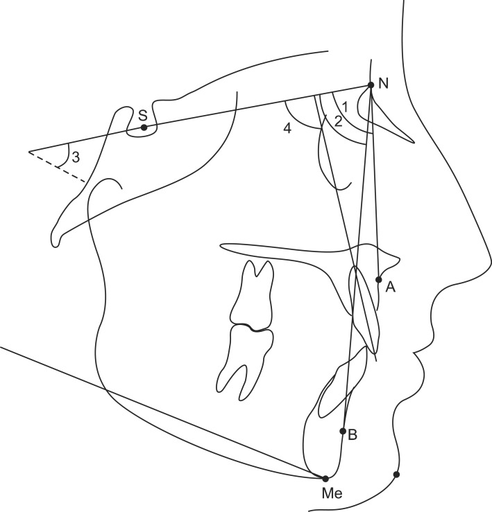
Figure 1 Landmarks and angular measurements on lateral cephalometric radiograph. The sella-nasion-A point (SNA) (1) angle relates to the anteroposterior position of the maxillary apical base to a line passing through the anterior cranial base. The sella-nasion-B point (SNB) (2) angle relates to the anteroposterior position of the mandibular apical base to a line passing through the anterior cranial base. The mandibular plane-anterior cranial base plane (sella-nasion/gonion-menton [SN-GoMe]) (3) angle relates to the cant of the mandibular plane to a line passing through the anterior cranial base. The maxillary incisor to anterior cranial base plane (1-SN) (4) angle relates to the axial inclination of the most labial maxillary incisor to a line passing through the anterior cranial base.

Figure 2 Landmarks and linear measurement on posteroanterior radiograph. Maxillary-maxillary apical base width (Mxr-Mxl) was defined as the horizontal distance between the right and left intersections of the lateral contour of the maxillary alveolar process and the lower contour of the zygomatic process of the maxilla.

Figure 3 Formation of the angles used for inclination assessment. α1 (right molar tipping angle) and α2 (left molar tipping angle), inner angles between the transversal occlusal line connecting the mesio-palatal cusp tips of the right and left molars and the lines passing through the mesio-buccal and mesio-palatal cusp tips of the molars. α3 (palatal tipping angle), inner angle between the right and left alveolar lines connecting the upper and lower alveolar tipping points on each side.
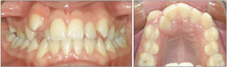
Figure 4 Pretreatment photographs of a patient.
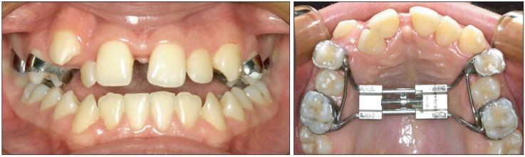
Figure 5 Photographs after the completion of maxillary expansion.
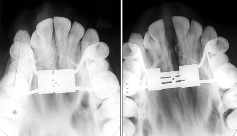
Figure 6 Occlusal radiograph showing sutural separation.
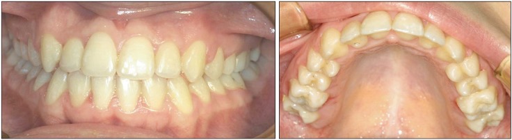
Figure 7 Post-treatment photographs.
Reference
-
1. Kurol J, Berglund L. Longitudinal study and cost-benefit analysis of the effect of early treatment of posterior cross-bites in the primary dentition. Eur J Orthod. 1992; 14:173–179. PMID: 1628683.
Article2. Lamparski DG Jr, Rinchuse DJ, Close JM, Sciote JJ. Comparison of skeletal and dental changes between 2-point and 4-point rapid palatal expanders. Am J Orthod Dentofacial Orthop. 2003; 123:321–328. PMID: 12637904.
Article3. Angell EC. Treatment of irregularities of the permanent or adult teeth. Dent Cosmos. 1860; 1:540–544.4. Haas AJ. Rapid expansion of the maxillary dental arch and nasal cavity by opening the midpalatal suture. Angle Orthod. 1961; 31:73–90.5. Isaacson RJ, Wood JL, Ingram AH. Forces produced by rapid maxillary expansion. I. Design of the force measuring system. Angle Orthod. 1964; 34:256–260.6. Isaacson RJ, Ingram AH. Forces produced by rapid maxillary expansion. II. Forces present during treatment. Angle Orthod. 1964; 34:261–270.7. Zimring JF, Isaacson RJ. Forces produced by rapid maxillary expansion. III. forces present during retention. Angle Orthod. 1965; 35:178–186. PMID: 14331018.8. Darendeliler MA, Strahm C, Joho JP. Light maxillary expansion forces with the magnetic expansion device. A preliminary investigation. Eur J Orthod. 1994; 16:479–490. PMID: 7720793.
Article9. Vardimon AD, Graber TM, Voss LR. Stability of magnetic versus mechanical palatal expansion. Eur J Orthod. 1989; 11:107–115. PMID: 2670588.
Article10. Harberson VA, Myers DR. Midpalatal suture opening during functional posterior cross-bite correction. Am J Orthod. 1978; 74:310–313. PMID: 362931.
Article11. Arndt WV. Nickel titanium palatal expander. J Clin Orthod. 1993; 27:129–137. PMID: 8496351.12. Wichelhaus A, Geserick M, Ball J. A new nickel titanium rapid maxillary expansion screw. J Clin Orthod. 2004; 38:677–680. PMID: 15665443.13. Oktay H, Kiliç N. Evaluation of the inclination in posterior dentoalveolar structures after rapid maxillary expansion: a new method. Dentomaxillofac Radiol. 2007; 36:356–359. PMID: 17699706.
Article14. Biederman W. Rapid correction of Class 3 malocclusion by midpalatal expansion. Am J Orthod. 1973; 63:47–55. PMID: 4565361.15. Kiliç N, Kiki A, Oktay H. A comparison of dentoalveolar inclination treated by two palatal expanders. Eur J Orthod. 2008; 30:67–72. PMID: 18276928.16. Haas AJ. Palatal expansion: just the beginning of dentofacial orthopedics. Am J Orthod. 1970; 57:219–255. PMID: 5263785.
Article17. Hicks EP. Slow maxillary expansion. A clinical study of the skeletal versus dental response to low-magnitude force. Am J Orthod. 1978; 73:121–141. PMID: 343597.18. Wertz RA. Skeletal and dental changes accompanying rapid midpalatal suture opening. Am J Orthod. 1970; 58:41–66. PMID: 5269181.
Article19. Mossaz-Joëlson K, Mossaz CF. Slow maxillary expansion: a comparison between banded and bonded appliances. Eur J Orthod. 1989; 11:67–76. PMID: 2653850.
Article20. Ciambotti C, Ngan P, Durkee M, Kohli K, Kim H. A comparison of dental and dentoalveolar changes between rapid palatal expansion and nickel-titanium palatal expansion appliances. Am J Orthod Dentofacial Orthop. 2001; 119:11–20. PMID: 11174532.
Article21. Sarver DM, Johnston MW. Skeletal changes in vertical and anterior displacement of the maxilla with bonded rapid palatal expansion appliances. Am J Orthod Dentofacial Orthop. 1989; 95:462–466. PMID: 2658544.
Article22. Krebs AA. Expansion of mid palatal suture studied by means of metallic implants. Acta Odontol Scand. 1959; 17:491–501.23. Sarnäs KV, Björk A, Rune B. Long-term effect of rapid maxillary expansion studied in one patient with the aid of metallic implants and roentgen stereometry. Eur J Orthod. 1992; 14:427–432. PMID: 1486927.
- Full Text Links
-
- Actions
-
Cited
- CITED
-
- Close
- Share
-
- Similar articles
-
- A posteroanterior cephalometric study on the change of maxilla by rapid palatal expansion
- A tomographic study on oro-nasal dimensional changes following rapid palatal expansion
- A study on the effect of rapid maxillary expansion and its relapse
- Salvage rapid maxillary expansion for the relapse of maxillary transverse expansion after Le Fort I with parasagittal osteotomy
- Crowding with no posterior crossbite treatment byrapid palatal expansion
