Surgery-first approach using a three-dimensional virtual setup and surgical simulation for skeletal Class III correction
- Affiliations
-
- 1Department of Orthodontics, National Health Insurance Service Ilsan Hospital, Goyang, Korea. kjunghoon@gmail.com
- 2Department of Oral and Maxillofacial Surgery, National Health Insurance Service Ilsan Hospital, Goyang, Korea.
- KMID: 2273233
- DOI: http://doi.org/10.4041/kjod.2014.44.6.330
Abstract
- A 19-year-old woman presented to our dental clinic with anterior crossbite and mandibular prognathism. She had a concave profile, long face, and Angle Class III molar relationship. She showed disharmony in the crowding of the maxillomandibular dentition and midline deviation. The diagnosis and treatment plan were established by a three-dimensional (3D) virtual setup and 3D surgical simulation, and a surgical wafer was produced using the stereolithography technique. No presurgical orthodontic treatment was performed. Using the surgery-first approach, Le Fort I maxillary osteotomy and mandibular bilateral intraoral vertical ramus osteotomy setback were carried out. Treatment was completed with postorthodontic treatment. Thus, symmetrical and balanced facial soft tissue and facial form as well as stabilized and well-balanced occlusion were achieved.
Keyword
MeSH Terms
Figure
-
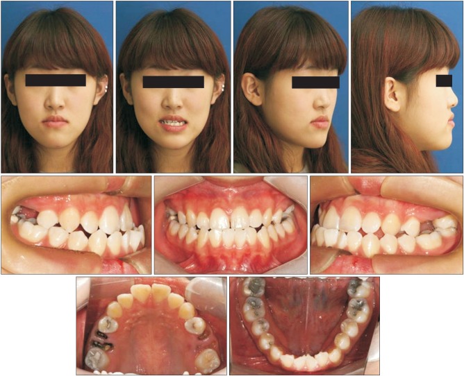
Figure 1 Pretreatment extraoral and intraoral photographs.

Figure 2 Pretreatment panoramic radiograph and lateral and frontal cephalometric radiographs.
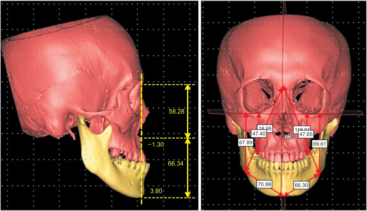
Figure 3 Pretreatment computed tomography images.
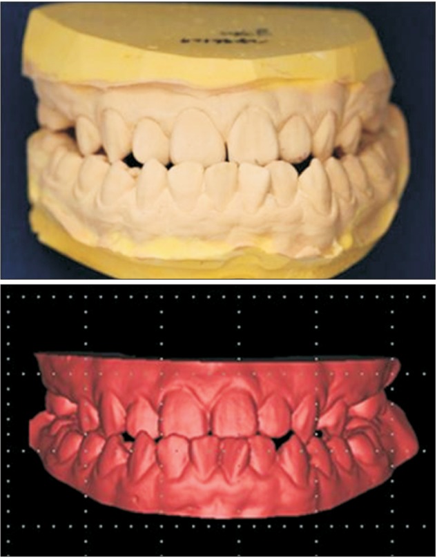
Figure 4 Initial dental cast model and three-dimensional digital model.
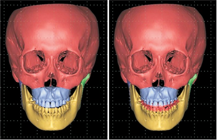
Figure 5 Superimposition of the initial digital cast model onto the three-dimensional computed tomography image.

Figure 6 Virtual setup based on prediction of the presurgical orthodontic treatment.

Figure 7 Three-dimensional surgical simulation using Mimics® 14.0 (Materialise, Leuven, Belgium).
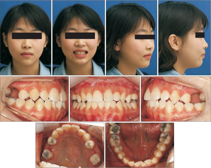
Figure 8 Postoperative extraoral and intraoral photographs.
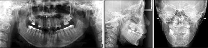
Figure 9 Postoperative panoramic radiograph and lateral and frontal cephalometric radiographs.

Figure 10 Intraoral photographs at one month after postsurgical orthodontic treatment.
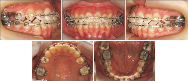
Figure 11 Intraoral photographs at 3 months after postsurgical orthodontic treatment.
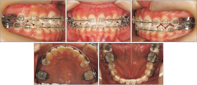
Figure 12 Intraoral photographs at 6 months after postsurgical orthodontic treatment.
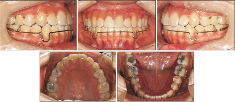
Figure 13 Clear retainer in the maxilla and circumferential retainer in the mandible.
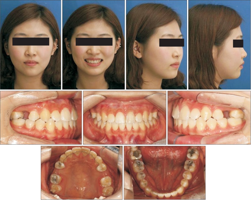
Figure 14 Posttreatment extraoral and intraoral photographs.
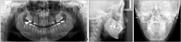
Figure 15 Posttreatment panoramic radiograph and lateral and frontal cephalometric radiographs.
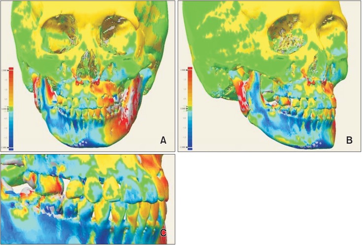
Figure 16 Comparison of the simulated cast and computed tomography (CT) images and the posttreatment CT images using superimposition and a color-coded discrepancy map. A, Frontal view; B, oblique view; C, dental view.
Cited by 1 articles
-
Evaluation of stability after pre-orthodontic orthognathic surgery using cone-beam computed tomography: A comparison with conventional treatment
Hye-Rim Ann, Young-Soo Jung, Kee-Joon Lee, Hyoung-Seon Baik
Korean J Orthod. 2016;46(5):301-309. doi: 10.4041/kjod.2016.46.5.301.
Reference
-
1. Dowling PA, Espeland L, Krogstad O, Stenvik A, Kelly A. Duration of orthodontic treatment involving orthognathic surgery. Int J Adult Orthodon Orthognath Surg. 1999; 14:146–152. PMID: 10686838.2. Luther F, Morris DO, Hart C. Orthodontic preparation for orthognathic surgery: how long does it take and why? A retrospective study. Br J Oral Maxillofac Surg. 2003; 41:401–406. PMID: 14614870.
Article3. Baek SH, Ahn HW, Kwon YH, Choi JY. Surgery-first approach in skeletal class III malocclusion treated with 2-jaw surgery: evaluation of surgical movement and postoperative orthodontic treatment. J Craniofac Surg. 2010; 21:332–338. PMID: 20186090.4. Hernández-Alfaro F, Guijarro-Martínez R, Molina-Coral A, Badía-Escriche C. "Surgery first" in bimaxillary orthognathic surgery. J Oral Maxillofac Surg. 2011; 69:e201–e207. PMID: 21470740.
Article5. Keim RG. Surgery-first orthognathics. J Clin Orthod. 2009; 43:77–78. PMID: 19276577.6. Liou EJ, Chen PH, Wang YC, Yu CC, Huang CS, Chen YR. Surgery-first accelerated orthognathic surgery: orthodontic guidelines and setup for model surgery. J Oral Maxillofac Surg. 2011; 69:771–780. PMID: 21257249.
Article7. Nagasaka H, Sugawara J, Kawamura H, Nanda R. "Surgery first" skeletal Class III correction using the Skeletal Anchorage System. J Clin Orthod. 2009; 43:97–105. PMID: 19276579.8. Sugawara J, Aymach Z, Nagasaka DH, Kawamura H, Nanda R. "Surgery first" orthognathics to correct a skeletal class II malocclusion with an impinging bite. J Clin Orthod. 2010; 44:429–438. PMID: 21038796.9. Yu CC, Chen PH, Liou EJ, Huang CS, Chen YR. A Surgery-first approach in surgical-orthodontic treatment of mandibular prognathism--a case report. Chang Gung Med J. 2010; 33:699–705. PMID: 21199616.10. Kang SH, Kim MK, Park SY, Lee JY, Park W, Lee SH. Early orthognathic surgery with three-dimensional image simulation during presurgical orthodontics in adults. J Craniofac Surg. 2011; 22:473–481. PMID: 21403556.
Article11. Sharifi A, Jones R, Ayoub A, Moos K, Walker F, Khambay B, et al. How accurate is model planning for orthognathic surgery? Int J Oral Maxillofac Surg. 2008; 37:1089–1093. PMID: 18760569.
Article12. Macchi A, Carrafiello G, Cacciafesta V, Norcini A. Three-dimensional digital modeling and setup. Am J Orthod Dentofacial Orthop. 2006; 129:605–610. PMID: 16679200.
Article13. Cevidanes LH, Tucker S, Styner M, Kim H, Chapuis J, Reyes M, et al. Three-dimensional surgical simulation. Am J Orthod Dentofacial Orthop. 2010; 138:361–371. PMID: 20816308.
Article14. Kang SH, Kim MK, Park WS, Lee SH. Accurate computerised mandibular simulation in orthognathic surgery: a new method for integrating the planned postoperative occlusion model. Br J Oral Maxillofac Surg. 2010; 48:305–307. PMID: 19616350.
Article15. Liou EJ, Chen PH, Wang YC, Yu CC, Huang CS, Chen YR. Surgery-first accelerated orthognathic surgery: postoperative rapid orthodontic tooth movement. J Oral Maxillofac Surg. 2011; 69:781–785. PMID: 21353934.
Article16. Villegas C, Uribe F, Sugawara J, Nanda R. Expedited correction of significant dentofacial asymmetry using a "surgery first" approach. J Clin Orthod. 2010; 44:97–103. PMID: 20552809.17. Frost HM. The regional acceleratory phenomenon: a review. Henry Ford Hosp Med J. 1983; 31:3–9. PMID: 6345475.18. Murphy KG, Wilcko MT, Wilcko WM, Ferguson DJ. Periodontal accelerated osteogenic orthodontics: a description of the surgical technique. J Oral Maxillofac Surg. 2009; 67:2160–2166. PMID: 19761909.
Article19. Wilcko MT, Wilcko WM, Pulver JJ, Bissada NF, Bouquot JE. Accelerated osteogenic orthodontics technique: a 1-stage surgically facilitated rapid orthodontic technique with alveolar augmentation. J Oral Maxillofac Surg. 2009; 67:2149–2159. PMID: 19761908.
Article20. Park SH, Yu HS, Kim KD, Lee KJ, Baik HS. A proposal for a new analysis of craniofacial morphology by 3-dimensional computed tomography. Am J Orthod Dentofacial Orthop. 2006; 129:600.e23–600.e34. PMID: 16679198.
Article21. Plooij JM, Maal TJ, Haers P, Borstlap WA, Kuijpers-Jagtman AM, Bergé SJ. Digital three-dimensional image fusion processes for planning and evaluating orthodontics and orthognathic surgery. A systematic review. Int J Oral Maxillofac Surg. 2011; 40:341–352. PMID: 21095103.
Article
- Full Text Links
-
- Actions
-
Cited
- CITED
-
- Close
- Share
-
- Similar articles
-
- Corrective Surgery Using Virtual Surgical Simulation and a Three-Dimensional Printed Osteotomy Guide: A Case Report
- The study of the soft tissue change according to skeletal change following bssro with advancing genioplasty
- Two treatment approach to skeletal Class III : A case report on sisters
- Surgery-first approach reduces the overall treatment time without damaging longterm stability in the skeletal class III correction: a preliminary study
- 2 Phase Treatment Without Preoperative Orthodontics In Skeletal Class III Malocclusion
