Comparison of condylar displacement between three biotypological facial groups by using mounted models and a mandibular position indicator
- Affiliations
-
- 1Department of Orthodontics, Faculty of Dental Medicine of University of Porto, Porto, Portugal. mponces@fmd.up.pt
- 2Department of Civil Engineering, Faculty of Engineering of University of Porto, Porto, Portugal.
- KMID: 2273231
- DOI: http://doi.org/10.4041/kjod.2014.44.6.312
Abstract
OBJECTIVE
Facial-type-associated variations in diagnostic features have several implications in orthodontics. For example, in hyperdivergent craniofacial types, growth imbalances are compensated by displacement of the condyle. When diagnosis and treatment planning involves centric relation (CR), detailed knowledge of the condylar position is desirable. The present study aimed to measure condylar displacement (CD) between CR and maximum intercuspation in three facial types of an asymptomatic orthodontic population.
METHODS
The study was conducted in 108 patients classified into three groups of 36 individuals each (27 women and 9 men; mean age, 20.5 years), based on the following facial patterns: hyperdivergent, hypodivergent, and intermediate. To quantify CD along the horizontal and vertical axes, the condylar position was analyzed using mounted casts on a semi-adjustable articulator and a mandibular position indicator. The Student t-test was used to compare CD between the groups.
RESULTS
Vertical displacement was found to be significantly different between the hyperdivergent and hypodivergent groups (p < 0.0002) and between the hyperdivergent and intermediate groups (p < 0.0006). The differences in horizontal displacement were not significant between the groups. In each group, vertical CD was more evident than horizontal displacement was.
CONCLUSIONS
All facial types, especially the hyperdivergent type, carried a significantly high risk of CD. Therefore, the possibility of CD should be carefully evaluated and considered in the assessment of all orthodontic cases in order to accurately assess jaw relationships and avoid possible misdiagnosis.
Keyword
MeSH Terms
Figure
-
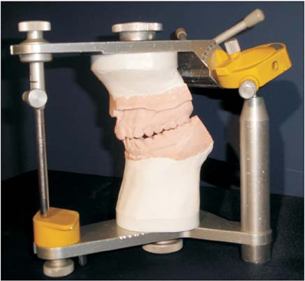
Figure 1 Study casts mounted in centric relation on an SAM® 2P semi-adjustable articulator.

Figure 2 A mandibular position indicator attached to a cast of the upper arch, and maximum intercuspation wax registration interposed between the two casts in order to register the corresponding condylar position.

Figure 3 A mandibular position indicator registration: black and red dots represent right and left condylar positions corresponding to centric relation and maximum intercuspation, respectively. Axial displacements are as follows: for the right condyle, -0.9 mm in XX' and +1.8 mm in ZZ'; and for the left condyle, -1.3 mm in XX' and +2.2 mm in ZZ'.
Cited by 1 articles
-
Three-dimensional cone-beam computed tomography based comparison of condylar position and morphology according to the vertical skeletal pattern
In-Young Park, Ji-Hyun Kim, Yang-Ho Park
Korean J Orthod. 2015;45(2):66-73. doi: 10.4041/kjod.2015.45.2.66.
Reference
-
1. Roth RH. Functional occlusion for the orthodontist. J Clin Orthod. 1981; 15:32–40. 44-51 contd.2. Shildkraut M, Wood DP, Hunter WS. The CR-CO discrepancy and its effect on cephalometric measurements. Angle Orthod. 1994; 64:333–342.3. Crawford SD. Condylar axis position, as determined by the occlusion and measured by the CPI instrument, and signs and symptoms of temporomandibular dysfunction. Angle Orthod. 1999; 69:103–115.4. Cordray FE. Three-dimensional analysis of models articulated in the seated condylar position from a deprogrammed asymptomatic population: a prospective study. Part 1. Am J Orthod Dentofacial Orthop. 2006; 129:619–630.
Article5. Rinchuse DJ, Kandasamy S. Myths of orthodontic gnathology. Am J Orthod Dentofacial Orthop. 2009; 136:322–330.
Article6. Isberg AM, Isacsson G. Tissue reactions of the temporomandibular joint following retrusive guidance of the mandible. Cranio. 1986; 4:143–148.
Article7. Slavicek R. Clinical and instrumental functional analysis and treatment planning. Part 4. Instrumental analysis of mandibular casts using the mandibular position indicator. J Clin Orthod. 1988; 22:566–575.8. Wood DP, Elliott RW. Reproducibility of the centric relation bite registration technique. Angle Orthod. 1994; 64:211–220.9. Beard CC, Clayton JA. Effects of occlusal splint therapy on TMJ dysfunction. J Prosthet Dent. 1980; 44:324–335.
Article10. Isaacson JR, Isaacson RJ, Speidel TM, Worms FW. Extreme variation in vertical facial growth and associated variation in skeletal and dental relations. Angle Orthod. 1971; 41:219–229.11. Hidaka O, Adachi S, Takada K. The difference in condylar position between centric relation and centric occlusion in pretreatment Japanese orthodontic patients. Angle Orthod. 2002; 72:295–301.12. Girardot RA Jr. Comparison of condylar position in hyperdivergent and hypodivergent facial skeletal types. Angle Orthod. 2001; 71:240–246.13. Desai S, Johnson DL, Howes RI, Rohrer MD. Changes in the rabbit temporomandibular joint associated with posterior displacement of the mandible. Int J Prosthodont. 1996; 9:46–57.14. Ahn SJ, Baek SH, Kim TW, Nahm DS. Discrimination of internal derangement of temporomandibular joint by lateral cephalometric analysis. Am J Orthod Dentofacial Orthop. 2006; 130:331–339.
Article15. Hwang CJ, Sung SJ, Kim SJ. Lateral cephalometric characteristics of malocclusion patients with temporomandibular joint disorder symptoms. Am J Orthod Dentofacial Orthop. 2006; 129:497–503.
Article16. Pullinger AG, Hollender L, Solberg WK, Petersson A. A tomographic study of mandibular condyle position in an asymptomatic population. J Prosthet Dent. 1985; 53:706–713.
Article17. Hatcher DC, Blom RJ, Baker CG. Temporomandibular joint spatial relationships: osseous and soft tissues. J Prosthet Dent. 1986; 56:344–353.
Article18. Ribeiro RF, Tallents RH, Katzberg RW, Murphy WC, Moss ME, Magalhaes AC, et al. The prevalence of disc displacement in symptomatic and asymptomatic volunteers aged 6 to 25 years. J Orofac Pain. 1997; 11:37–47.19. Ikeda K, Kawamura A. Assessment of optimal condylar position with limited cone-beam computed tomography. Am J Orthod Dentofacial Orthop. 2009; 135:495–501.
Article20. Cordray FE. The importance of the seated condylar position in orthodontic correction. Quintessence Int. 2002; 33:284–293.21. Alexander SR, Moore RN, DuBois LM. Mandibular condyle position: comparison of articulator mountings and magnetic resonance imaging. Am J Orthod Dentofacial Orthop. 1993; 104:230–239.
Article22. Levine D, Gosink BB. Ultrasound shows changes in postmenopausal pelvis. Diagn Imaging (San Franc). 1992; 14:96–101.23. Karl PJ, Foley TF. The use of a deprogramming appliance to obtain centric relation records. Angle Orthod. 1999; 69:117–124.24. Burke G, Major P, Glover K, Prasad N. Correlations between condylar characteristics and facial morphology in Class II preadolescent patients. Am J Orthod Dentofacial Orthop. 1998; 114:328–336.
Article25. Helkimo M. Epidemiological surveys of dysfunction of the masticatory system. Oral Sci Rev. 1976; 7:54–69.26. Fantini SM, Paiva JB, Rino Neto J, Dominguez GC, Abrão J, Vigoritto JW. Increase of condylar displacement between centric relation and maximal habitual intercuspation after occlusal splint therapy. Braz Oral Res. 2005; 19:176–182.
Article27. Baccetti T, Franchi L, McNamara JA Jr. The Cervical Vertebra Maturation (CVM) method for the assessment of the optimal treatment timing in dentofacial orthopedics. Semin Orthod. 2005; 11:119–129.
Article28. Ramalhão MJ. Deslocamento condilar nos tipos faciais hiperdivergentes [Thesis]. Porto: University of Porto;2009.29. Wood DP, Floreani KJ, Galil KA, Teteruck WR. The effect of incisal bite force on condylar seating. Angle Orthod. 1994; 64:53–61.30. McNamara JA Jr, Seligman DA, Okeson JP. Occlusion, Orthodontic treatment, and temporomandibular disorders: a review. J Orofac Pain. 1995; 9:73–90.
- Full Text Links
-
- Actions
-
Cited
- CITED
-
- Close
- Share
-
- Similar articles
-
- Is distal segment ostectomy essential for stabilization of the condylar position in patients with facial asymmetry?
- Positional change in mandibular condyle in facial asymmetric patients after orthognathic surgery: cone-beam computed tomography study
- Three-dimensional cone-beam computed tomography based comparison of condylar position and morphology according to the vertical skeletal pattern
- The relationship of mandibular condylar position to overbite depth
- The study of the effect of mandibular growth and function in pediatric unilateral condyle fractures
