Korean J Orthod.
2010 Oct;40(5):334-341. 10.4041/kjod.2010.40.5.334.
Comparison of arch form between Koreans and Egyptians
- Affiliations
-
- 1Department of Orthodontics, Graduate School of Clinical Dental Science, The Catholic University of Korea, Seoul, Korea.
- 2Department of Orthodontics, School of Dentistry, Kyunghee University, Seoul, Korea.
- 3Department of Orthodontics, Seoul St. Mary's Hospital, The Catholic University of Korea, Seoul, Korea. kook2002@catholic.ac.kr
- KMID: 2273224
- DOI: http://doi.org/10.4041/kjod.2010.40.5.334
Abstract
OBJECTIVE
The purpose of this study was to evaluate morphologic differences in the mandibular arch between Egyptian and Korean subjects.
METHODS
The Egyptian sample consisted of 94 mandibular casts (35 Class I, 32 Class II and 27 Class III). The Korean sample consisted of 462 mandibular casts (114 Class I, 119 Class II, and 135 Class III). The most facial portion of 13 proximal contact areas was digitized from photocopied images of the mandibular dental arches. Clinical bracket points were calculated for each tooth. The subjects were grouped according to arch form to compare the frequency distribution of the 3 arch forms between the ethnic groups in each Angle classification.
RESULTS
Egyptians had significantly narrower intermolar and intercanine widths (p < 0.001), and shallower intermolar and intercanine depths (p < 0.001) than Koreans. There was an even frequency distribution of the 3 arch forms within the Egyptian group (p = 0.46). However, in the Korean group, the most frequent arch form was the square arch form (46.7%), while the frequency of the tapered arch form was significantly lower (18.8%).
CONCLUSIONS
These results might provide helpful information in evaluating morphologic differences between ethnic groups in selection of preformed superelastic archwires.
Keyword
MeSH Terms
Figure
-
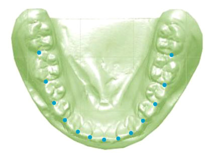
Fig. 1 Digitized contact points on a mandibular cast.

Fig. 2 A perpendicular line extended from a line connecting the contact points to find the clinical bracket points.
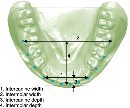
Fig. 3 Twelve bracket points and 4 linear measurements.
Cited by 1 articles
-
Comparison of arch form between ethnic Malays and Malaysian Aborigines in Peninsular Malaysia
Siti Adibah Othman, Eunice Soh Xinwei, Sheh Yinn Lim, Marhazlinda Jamaludin, Nor Himazian Mohamed, Zamros Yuzaidi Mohd Yusof, Lily Azura Shoaib, Nik Noriah Nik Hussein
Korean J Orthod. 2012;42(1):47-54. doi: 10.4041/kjod.2012.42.1.47.
Reference
-
1. de la Cruz A, Sampson P, Little RM, Artun J, Shapiro PA. Long-term changes in arch form after orthodontic treatment and retention. Am J Orthod Dentofacial Orthop. 1995. 107:518–530.
Article2. Felton JM, Sinclair PM, Jones DL, Alexander RG. A computerized analysis of the shape and stability of mandibular arch form. Am J Orthod Dentofacial Orthop. 1987. 92:478–483.
Article3. McLaughlin RP, Bennett JC. Arch form considerations for stability and esthetics. Rev Esp Orthod. 2000. 29:46–63.4. Shapiro PA. Mandibular dental arch form and dimension: Treatment and postretention changes. Am J Orthod. 1974. 66:58–70.5. Krishnan M, Kalathil S, Abraham KM. Comparative evaluation of frictional forces in active and passive self-ligating brackets with various archwire alloys. Am J Orthod Dentofacial Orthop. 2009. 136:675–682.
Article6. Ferrario VF, Sforza C, Miani A Jr, Tartaglia G. Mathematical definition of the shape of dental arches in human permanent healthy dentitions. Eur J Orthod. 1994. 16:287–294.
Article7. Merz ML, Isaacson RJ, Germane N, Rubenstein LK. Tooth diameters and arch perimeters in a black and a white population. Am J Orthod Dentofacial Orthop. 1991. 100:53–58.
Article8. Lee CH, Mo SS, Kang YG, Nojima K, Kim YH, Kook YA. Comparison of arch forms between Korean and Japanese in Class I, II, and III malocclusion. Korean J Orthod. 2007. 37:364–375.9. Kunjur J, Sabesan T, Ilankovan V. Anthropometric analysis of eyebrows and eyelids: an inter-racial study. Br J Oral Maxillofac Surg. 2006. 44:89–93.
Article10. Behbehani F, Hicks EP, Beeman C, Kluemper GT, Rayens MK. Racial variations in cephalometric analysis between Whites and Kuwaitis. Angle Orthod. 2006. 76:406–411.11. Lee JJ, Ramirez SG, Will MJ. Gender and racial variations in cephalometric analysis. Otolaryngol Head Neck Surg. 1997. 117:326–329.
Article12. Abd-el Samad Younes S. Maxillary arch dimensions in Saudi and Egyptian population sample. Am J Orthod. 1984. 85:83–88.
Article13. Bishara SE, Abdalla EM, Hoppens BJ. Cephalometric comparisons of dentofacial parameters between Egyptian and North American adolescents. Am J Orthod Dentofacial Orthop. 1990. 97:413–421.
Article14. Sarhan OA, Diwan RR. Maxillary arch dimensions in Egyptian and British children. Odontostomatol Trop. 1987. 10:101–106.15. Kook YA, Nojima K, Moon HB, McLaughlin RP, Sinclair PM. Comparison of arch forms between Korean and North American white populations. Am J Orthod Dentofacial Orthop. 2004. 126:680–686.
Article16. The World Factbook, Egypt [Internet]. Available from: http://www.cia.gov/library/publications/the-world-factbook/print/eg.html.17. Arab League. Wikipedia, the free encyclopedia [Internet]. Available from: http://en.wikipedia.org/wiki/Arab_League.18. Andrews LF. Straight wire - the concept and appliance. 1989. San Diego: LA Wells.19. Nummikoski P, Prihoda T, Langlais RP, McDavid WD, Welander U, Tronje G. Dental and mandibular arch widths in three ethnic groups in Texas: a radiographic study. Oral Surg Oral Med Oral Pathol. 1988. 65:609–617.
Article20. Collins BP, Harris EF. Arch form in American blacks and whites with malocclusions. J Tenn Dent Assoc. 1998. 78:15–18.21. Burris BG, Harris EF. Maxillary arch size and shape in American blacks and whites. Angle Orthod. 2000. 70:297–302.22. BeGole EA. Application of the cubic spline function in the description of dental arch form. J Dent Res. 1980. 59:1549–1556.
Article23. Bonwill WGA. Geometrical and mechanical laws of articulation. Trans Odont Soc Penn. 1884-1885. 119–133.24. Camporesi M, Franchi L, Baccetti T, Antonini A. Thin-plate spline analysis of arch form in a Southern European population with an ideal natural occlusion. Eur J Orthod. 2006. 28:135–140.
Article25. Ferrario VF, Sforza C, Miani A Jr, Tartaglia G. Maxillary versus mandibular arch form differences in human permanent dentition assessed by Euclidean-distance matrix analysis. Arch Oral Biol. 1994. 39:135–139.
Article26. Noroozi H, Nik TH, Saeeda R. The dental arch form revisited. Angle Orthod. 2001. 71:386–389.27. Taner TU, Ciger S, El H, Germeç D, Es A. Evaluation of dental arch width and form changes after orthodontic treatment and retention with a new computerized method. Am J Orthod Dentofacial Orthop. 2004. 126:464–475.
Article28. Nojima K, McLaughlin RP, Isshiki Y, Sinclair PM. A comparative study of Caucasian and Japanese mandibular clinical arch forms. Angle Orthod. 2001. 71:195–200.29. DeKock WH. Dental arch depth and width studied longitudinally from 12 years of age to adulthood. Am J Orthod. 1972. 62:56–66.
Article30. Haralabakis NB, Sifakakis I, Papagrigorakis M, Papadakis G. The correlation of sexual dimorphism in tooth size and arch form. World J Orthod. 2006. 7:254–260.31. Braun S, Hnat WP, Fender DE, Legan HL. The form of the human dental arch. Angle Orthod. 1998. 68:29–36.32. Kook YA, Bayome M, Park SB, Cha BK, Lee YW, Baek SH. Overjet at the anterior and posterior segments: three-dimensional analysis of arch coordination. Angle Orthod. 2009. 79:495–501.
Article
- Full Text Links
-
- Actions
-
Cited
- CITED
-
- Close
- Share
-
- Similar articles
-
- An experimental study of ideal arch form of the Class I malocclusion patients with first premolar extraction
- A study on the dental arch by occlusogram in normal occlusion
- The dental arch form in normal occlusion
- Mandibular clinical arch forms in Koreans with normal occlusions
- A comparison of the form of the face, the dental arch and the maxillary central incisor
