Korean J Orthod.
2010 Apr;40(2):106-114. 10.4041/kjod.2010.40.2.106.
Stimulation of bone formation by direct electrical current in an orthopedically expanded suture in the rat
- Affiliations
-
- 1Department of Orthodontics, Faculty of Dentistry, Erciyes University, Kayseri Turkey, and Visiting Professor, King Saud University, Riyadh, Saudi Arabia. tancanuysal@yahoo.com
- 2Department of Orthodontics, Centre of Dental Sciences, Guhane Military Medical Academy, Ankara, Turkey.
- 3Department of Pathology, Faculty of Medicine, Guhane Military Medical Academy, Ankara, Turkey.
- KMID: 2273218
- DOI: http://doi.org/10.4041/kjod.2010.40.2.106
Abstract
OBJECTIVE
The aim of this experimental study was to evaluate the effects of direct electrical current stimulation (DECS) on bone regeneration in response to an expansion of the inter-premaxillary suture in the rat.
METHODS
Sixteen 50 - 60 days old Wistar male rats were separated into two equal groups (control and experimental). Both groups were subjected to expansion, and 30-gram of force was applied to the maxillary incisors with helical-spring. In the experimental group, two metallic-screws were placed at lateral parts of the maxillary segments. Electrodes were connected to the screws. The device was activated with current adjustment to measure 10 microA continuously and the current was monitored daily during the expansion and early-retention phase. Bone regeneration in the sutural area was histomorphometrically evaluated including new-bone area (micrometer2), bone perimeter (micrometer), feret's diameter (micrometer) and newly formed bone (%) parameters. Kruskal-Wallis rank and Mann-Whitney U tests were used for statistical evaluation at p < 0.05 level.
RESULTS
Statistical analysis showed significant differences between groups for all investigated histomorphometric parameters. New bone area (p = 0.002), bone perimeter (p = 0.004), feret's diameter (p = 0.002) and newly formed bone percentage (p = 0.002) measurements were significantly higher in the experimental group than the control group. Bone histomorphometric measurements revealed that bone architecture in the DECS group was improved.
CONCLUSIONS
The application of DECS to an orthopedically expanded inter-premaxillary suture area during the early retention phase stimulated the formation of new bone.
MeSH Terms
Figure
-

Fig. 1 Appliance in situ.
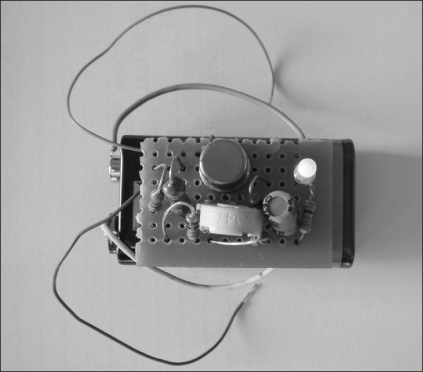
Fig. 2 Direct current electrical stimulator.
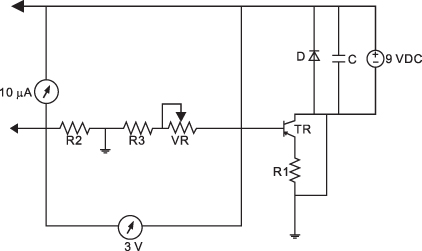
Fig. 3 Schematic diagram of the electrical circuit used to deliver 10 µA.
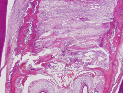
Fig. 4 Histological section of an expanded suture (H&E, ×40 magnification).

Fig. 5 Histomorphometric measurements of newly formed bone area (µm2). Image enhancement and outlining of the area of interest, which is newly formed bone.
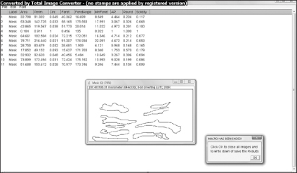
Fig. 6 At the end of the first macro, results window shows the basic planimetric measurements calculated from the outlined objects.

Fig. 7 Histomorphometric measurements of newly formed bone (percentage). The number of grid intersections on new bone and non-osseous connective tissue were counted and the percentage of new bone calculated.
Reference
-
1. Choi IH, Chung CY, Cho TJ, Yoo WJ. Angiogenesis and mineralization during distraction osteogenesis. J Korean Med Sci. 2002. 17:435–447.
Article2. Krebs AA. Midpalatal suture expansion studied by the implant method over a seven-year period. Trans Eur Orthod Soc. 1964. 40:131–142.3. Vardimon AD, Graber TM, Voss LR. Stability of magnetic versus mechanical palatal expansion. Eur J Orthod. 1989. 11:107–115.
Article4. Timms DJ. Long term follow-up of cases treated by rapid maxillary expansion. Trans Eur Orthod Soc. 1976. 52:211–215.5. Haas AJ. The treatment of maxillary deficiency by opening the midpalatal suture. Angle Orthod. 1965. 35:200–217.6. Saito S, Shimizu N. Stimulatory effects of low-power laser irradiation on bone regeneration in midpalatal suture during expansion in the rat. Am J Orthod Dentofacial Orthop. 1997. 111:525–532.
Article7. Sawada M, Shimizu N. Stimulation of bone formation in the expanding mid-palatal suture by transforming growth factor-beta 1 in the rat. Eur J Orthod. 1996. 18:169–179.
Article8. Uysal T, Ustdal A, Sonmez MF, Ozturk F. Stimulation of bone formation by dietary boron in an orthopedically expanded suture in rabbits. Angle Orthod. 2009. 79:984–990.
Article9. Uysal T, Amasyali M, Enhos S, Sonmez MF, Sagdic D. Effect of ED-71, a new active vitamin D analog, on bone formation in an orthopedically expanded suture in rats. A histomorphometric study. Eur J Dent. 2009. 3:165–172.
Article10. Uysal T, Amasyali M, Olmez H, Karslioglu Y, Gunhan O. Stimulation of bone formation in the expanding inter-premaxillary suture by vitamin E, in rat. Korean J Orthod. 2009. 39:337–347.
Article11. Albert SF, Wong E. Electrical stimulation of bone repair. Clin Podiatr Med Surg. 1991. 8:923–935.12. Blank M. Blank M, Findl E, editors. The influence of surface charge on oligomeric reactions as a basis for channel dynamics. Mechanistic Approaches to interactions of electric and electromagnetic fields with living systems. 1987. New York: Plenum Press;151–160.
Article13. Block MS, Brister GD. Use of distraction osteogenesis for maxillary advancement: preliminary results. J Oral Maxillofac Surg. 1994. 52:282–286.
Article14. Brighton CT, Black J, Friedenberg ZB, Esterhai JL, Day LJ, Connolly JF. A multicenter study of the treatment of non-union with constant direct current. J Bone Joint Surg Am. 1981. 63:2–13.
Article15. Cane V, Zaffe D, Cavani F, Botti P, Soana S. PEMFs modulate the enzymatic activity during the bone repair process. Bone. 1996. 19:133S.
Article16. Kim DH, Park YG, Kang SG. The effects of electrical current from a micro-electrical device on tooth movement. Korean J Orthod. 2008. 38:337–346.
Article17. El-Hakim IE, Azim AM, El-Hassan MF, Maree SM. Preliminary investigation into the effects of electrical stimulation on mandibular distraction osteogenesis in goats. Int J Oral Maxillofac Surg. 2004. 33:42–47.
Article18. Hagiwara T, Bell WH. Effect of electrical stimulation on mandibular distraction osteogenesis. J Craniomaxillofac Surg. 2000. 28:12–19.
Article19. Rasband WS. Image-J. 1997-2008. Bethesda, Maryland, USA: U.S. National Institutes of Health; http://rsb.info.nih.gov/ij/.20. Swennen G, Dempf R, Schliephage H. Cranio-facial distraction osteogenesis: a review of the literature. Part II: experimental studies. Int J Oral Maxillofac Surg. 2002. 31:123–135.
Article21. Storey E. Tissue response to the movement of bones. Am J Orthod. 1973. 64:229–247.
Article22. Burstone CJ, Shafer WG. Sutural expansion by controlled mechanical stress in the rat. J Dent Res. 1959. 38:534–540.
Article23. Cope JB, Samchukov ML. Mineralization dynamics of regenerate bone during mandibular osteodistraction. Int J Oral Maxillofac Surg. 2001. 30:234–242.
Article24. Matsunaga S. Histological, histochemical investigations of constant direct current stimulated intramedullary callus. Nippon Seikeigeka Gakkai Zasshi. 1986. 60:1293–1303.
- Full Text Links
-
- Actions
-
Cited
- CITED
-
- Close
- Share
-
- Similar articles
-
- EFFECT OF ELECTRICAL STIMULATION ON BONE FORMATION IN THE EXTRACTION SOCKET OF RAT
- Clinical Application of Direct Current Stimulation in the Treatment of Infected Non-Unions
- The Effect of Electrical Stimulation on Bone: An Experimental Study on Rabbits
- The Effect of Duration of Electrical Stimulation on New Bone Formation
- Stimulation of bone formation in the expanding inter-premaxillary suture by vitamin E, in rat
