Cone-beam computed tomography assessment of mandibular asymmetry in unilateral cleft lip and palate patients
- Affiliations
-
- 1Department of Orthodontics, Faculty of Dentistry, Dicle University, Diyarbakir, Turkey.
- 2Department of Orthodontics, Faculty of Dentistry, Izmir Katip Celebi University, Izmir, Turkey. tancan.uysal@ikc.edu.tr
- 3Department of Pediatric Dentistry and Orthodontics, College of Dentistry, King Saud University, Riyadh, Saudi Arabia.
- 4Department of Orthodontics, Faculty of Dentistry, Erciyes University, Kayseri, Turkey.
- KMID: 2273191
- DOI: http://doi.org/10.4041/kjod.2011.41.6.431
Abstract
OBJECTIVE
To determine whether there is any difference between the cleft and non-cleft sides of the mandible in unilateral cleft lip and palate (UCLP) patients, or the right and left sides in control patients; and to determine if there is any difference between the mandibular asymmetry of UCLP patients and that of control patients.
METHODS
We examined cone-beam computed tomography (CBCT) scans of 15 patients with UCLP and 15 age- and gender-matched control patients. We evaluated 8 linear, 3 surface, and 3 volumetric measurements and compared the cleft/non-cleft sides of UCLP patients and the right/left sides of controls.
RESULTS
There were no statistically significant gender differences in any linear, surface, or volumetric measurement. The single significant side-to-side difference in UCLP patients was a longer coronoid unit on the cleft side than on the non-cleft side (p = 0.046). Body volume was significantly lower in the UCLP group than in the control group (p = 0.008).
CONCLUSIONS
In general, UCLP patients have symmetrical mandibles, although the coronoid unit length is significantly longer on the cleft side than on the non-cleft side. UCLP patients and controls differed only in body volume.
Figure
-
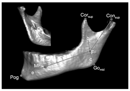
Fig. 1 Landmarks and measurements used in this study. A, condylar unit length; b, coronoid unit length; c, angular unit length; d, body unit length; e, chin unit length; Corsup, coronoid superius; Consup, condylion superius; F, fossa of mandibular foramen; Gomid, gonion midpoint; MF, mental foramen; Pog, pogonion.

Fig. 2 Landmarks and measurements used in this study. f, Condylar width; g, ramal height; h, body length; Jlat, the most lateral and deepest point of the curvature formed at the junction of the mandibular ramus and body; Jmed, The most medial and deepest point of the curvature formed at the junction of the mandibular ramus and body; Gomid, gonion midpoint; Consup, condylion superius; Conmed, condylion medialis; Conlat, condylion lateralis; Me, menton.

Fig. 3 An example of surface and volumetric measurements.
Cited by 1 articles
-
Relationship between chin deviation and the position and morphology of the mandible in individuals with a unilateral cleft lip and palate
Kyung-Seon Kim, Woo-Sung Son, Soo-Byung Park, Seong-Sik Kim, Yong-Il Kim
Korean J Orthod. 2013;43(4):168-177. doi: 10.4041/kjod.2013.43.4.168.
Reference
-
1. Liukkonen M, Sillanmäki L, Peltomäki T. Mandibular asymmetry in healthy children. Acta Odontol Scand. 2005. 63:168–172.
Article2. Pirttiniemi P, Raustia A, Kantomaa T, Pyhtinen J. Relationships of bicondylar position to occlusal asymmetry. Eur J Orthod. 1991. 13:441–445.
Article3. Van Elslande DC, Russett SJ, Major PW, Flores-Mir C. Mandibular asymmetry diagnosis with panoramic imaging. Am J Orthod Dentofacial Orthop. 2008. 134:183–192.
Article4. Bishara SE, Burkey PS, Kharouf JG. Dental and facial asymmetries: a review. Angle Orthod. 1994. 64:89–98.5. Laspos CP, Kyrkanides S, Tallents RH, Moss ME, Subtelny JD. Mandibular asymmetry in noncleft and unilateral cleft lip and palate individuals. Cleft Palate Craniofac J. 1997. 34:410–416.
Article6. Smahel Z, Brejcha M. Differences in craniofacial morphology between complete and incomplete unilateral cleft lip and palate in adults. Cleft Palate J. 1983. 20:113–127.7. Kurt G, Bayram M, Uysal T, Ozer M. Mandibular asymmetry in cleft lip and palate patients. Eur J Orthod. 2010. 32:19–23.
Article8. Horswell BB, Levant BA. Craniofacial growth in unilateral cleft lip and palate: skeletal growth from eight to eighteen years. Cleft Palate J. 1988. 25:114–121.9. Persson M. Mandibular asymmetry of hereditary origin. Am J Orthod. 1973. 63:1–11.
Article10. Schmid W, Mongini F, Felisio A. A computer-based assessment of structural and displacement asymmetries of the mandible. Am J Orthod Dentofacial Orthop. 1991. 100:19–34.
Article11. Hwang HS, Hwang CH, Lee KH, Kang BC. Maxillofacial 3-dimensional image analysis for the diagnosis of facial asymmetry. Am J Orthod Dentofacial Orthop. 2006. 130:779–785.
Article12. Van Elslande DC, Russett SJ, Major PW, Flores-Mir C. Mandibular asymmetry diagnosis with panoramic imaging. Am J Orthod Dentofacial Orthop. 2008. 134:183–192.
Article13. Ziegler CM, Woertche R, Brief J, Hassfeld S. Clinical indications for digital volume tomography in oral and maxillofacial surgery. Dentomaxillofac Radiol. 2002. 31:126–130.
Article14. Scarfe WC, Farman AG, Sukovic P. Clinical applications of cone-beam computed tomography in dental practice. J Can Dent Assoc. 2006. 72:75–80.15. Kyrkanides S, Klambani M, Subtelny JD. Cranial base and facial skeleton asymmetries in individuals with unilateral cleft lip and palate. Cleft Palate Craniofac J. 2000. 37:556–561.
Article16. Steiner CC. Cephalometrics in clinical practice. Angle Orthod. 1959. 29:8–29.17. Grummons DC, Kappeyne van de Coppello MA. A frontal asymmetry analysis. J Clin Orthod. 1987. 21:448–465.18. You KH, Lee KJ, Lee SH, Baik HS. Three-dimensional computed tomography analysis of mandibular morphology in patients with facial asymmetry and mandibular prognathism. Am J Orthod Dentofacial Orthop. 2010. 138:540.e1–540.e8.
Article19. Park W, Kim BC, Yu HS, Yi CK, Lee SH. Architectural characteristics of the normal and deformity mandible revealed by three-dimensional functional unit analysis. Clin Oral Investig. 2009. 14:691–698.
Article20. Kyrkanides S, Richter L. Mandibular asymmetry and antigonial notching in individuals with unilateral cleft lip and palate. Cleft Palate Craniofac J. 2002. 39:30–35.
Article21. Severt TR, Proffit WR. The prevalence of facial asymmetry in the dentofacial deformities population at the University of North Carolina. Int J Adult Orthodon Orthognath Surg. 1997. 12:171–176.22. Maeda M, Katsumata A, Ariji Y, Muramatsu A, Yoshida K, Goto S, et al. 3D-CT evaluation of facial asymmetry in patients with maxillofacial deformities. Oral Surg Oral Med Oral Pathol Oral Radiol Endod. 2006. 102:382–390.
Article23. Kyrkanides S, Bellohusen R, Subtelny JD. Skeletal asymmetries of the nasomaxillary complex in noncleft and postsurgical unilateral cleft lip and palate individuals. Cleft Palate Craniofac J. 1995. 32:428–433.
Article24. da Silva Filho OG, Normando AD, Capelozza Filho L. Mandibular growth in patients with cleft lip and/or cleft palate--the influence of cleft type. Am J Orthod Dentofacial Orthop. 1993. 104:269–275.
Article25. Krogman WM, Jain RB, Oka SW. Craniofacial growth in different cleft types from one month to ten years. Cleft Palate J. 1982. 19:206–211.26. Laspos CP, Kyrkanides S, Tallents RH, Moss ME, Subtelny JD. Mandibular and maxillary asymmetry in individuals with unilateral cleft lip and palate. Cleft Palate Craniofac J. 1997. 34:232–239.
Article27. Athanasiou AE, Moyers RE, Mazaheri M, Toutountzakis N. Frontal cephalometric evaluation of transverse dentofacial morphology and growth of children with isolated cleft palate. J Craniomaxillofac Surg. 1991. 19:249–253.
Article28. Moss ML, Rankow RM. The role of the functional matrix in mandibular growth. Angle Orthod. 1968. 38:95–103.29. Kwon TG, Lee KH, Park HS, Ryoo HM, Kim HJ, Lee SH. Relationship between the masticatory muscles and mandibular skeleton in mandibular prognathism with and without asymmetry. J Oral Maxillofac Surg. 2007. 65:1538–1543.
Article30. Smahel Z, Müllerová Z. Craniofacial morphology in unilateral cleft lip and palate prior to palatoplasty. Cleft Palate J. 1986. 23:225–232.31. Horswell BB, Levant BA. Craniofacial growth in unilateral cleft lip and palate: skeletal growth from eight to eighteen years. Cleft Palate J. 1988. 25:114–121.32. Lascala CA, Panella J, Marques MM. Analysis of the accuracy of linear measurements obtained by cone beam computed tomography (CBCT-NewTom). Dentomaxillofac Radiol. 2004. 33:291–294.
Article
- Full Text Links
-
- Actions
-
Cited
- CITED
-
- Close
- Share
-
- Similar articles
-
- Relationship between chin deviation and the position and morphology of the mandible in individuals with a unilateral cleft lip and palate
- Three-dimensional evaluation of midfacial asymmetry in patients with nonsyndromic unilateral cleft lip and palate by cone-beam computed tomography
- A cephalometric study on the position of the hyoid bone in cleft lip and palate individuals
- Facial asymmetry of unilateral cleft lip and palate patients
- A study on the maxillary dental arch and palate of unilateral cleft lip and palate individuals
