Relationship between chin deviation and the position and morphology of the mandible in individuals with a unilateral cleft lip and palate
- Affiliations
-
- 1Department of Orthodontics, School of Dentistry, Pusan National University, Yangsan, Korea. wsson@pusan.ac.kr
- KMID: 2272252
- DOI: http://doi.org/10.4041/kjod.2013.43.4.168
Abstract
OBJECTIVE
In this study, we aimed to examine the relationship between chin deviation and the positional and morphological features of the mandible and to determine the factors that contributed to chin deviation in individuals with a unilateral cleft lip and palate (UCLP).
METHODS
Cone-beam computed tomography (CBCT) images of 28 adults with UCLP were analyzed in this study. Segmented three-dimensional temporomandibular fossa and mandible images were reconstructed, and angular, linear, and volumetric parameters were measured.
RESULTS
For all 28 individuals, the chin was found to deviate to the cleft side by 1.59 mm. Moreover, among these 28 individuals, only 7 showed distinct (more than 4 mm) chin deviation, which was toward the cleft side. Compared to the non-cleft side, the mandibular body length, frontal ramal inclination, and vertical position of the condyle were lower and inclination of the temporomandibular fossa was steeper on the cleft side. Furthermore, the differences in inclination of the temporomandibular fossa, mandibular body length, ramus length, and condylar volume ratio (non-deviated/deviated) were positively correlated with chin deviation.
CONCLUSIONS
UCLP individuals show mild chin deviation to the cleft side. Statistical differences were noted in the parameters that represented positional and morphological asymmetries of the mandible and temporomandibular fossa; however, these differences were too small to indicate clinical significance.
Keyword
Figure
-
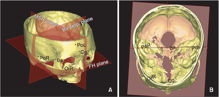
Figure 1 Three-dimensional reference planes used in this study. A, FH plane and MSR plane. B, Ba-perp. plane. PoL, Left ponon; PoR, right ponon; OrR, right orbitale. See Table 2 for the definitions of all other points and planes.
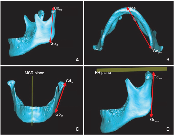
Figure 2 Three-dimensional measurements of mandible. A, Ramus length. B, Mandibular body length. C, Frontal ramal inclination. D, Lateral ramal inclination. See Table 2 for the definitions of all the points, planes, and measurements.
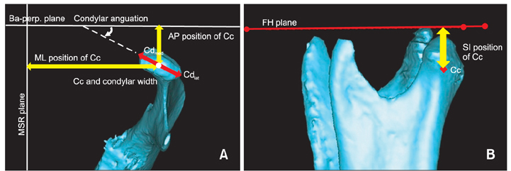
Figure 3 Measurement of the mandibular condyle. A, AP and ML positions of the Cc, condylar width, and condylar angulation. B, SI position of Cc. See Tables 2 and 3 for the definitions of all the points, planes, and measurements.
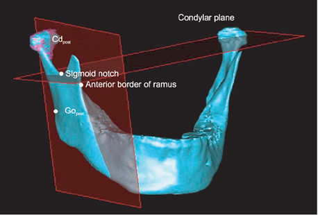
Figure 4 Segmentation of the mandibular condyle for condylar measurements. See Tables 2 and 3 for the definitions of all the points, planes, and measurements.
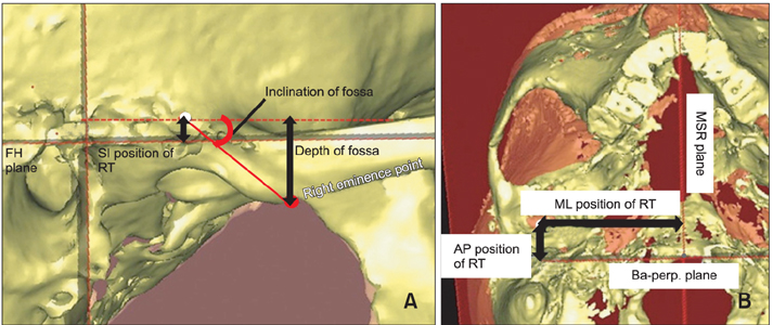
Figure 5 Three-dimensional measurements of the temporomandibular fossa. A, SI position of the RT, inclination of the temporomandibular fossa, and depth of the temporomandibular fossa. B, AP and ML positions of RT. See Tables 2 and 3 for the definitions of all the points, planes, and measurements.
Reference
-
1. Shetye PR, Evans CA. Midfacial morphology in adult unoperated complete unilateral cleft lip and palate patients. Angle Orthod. 2006; 76:810–816.2. Mølsted K, Dahl E. Asymmetry of the maxilla in children with complete unilateral cleft lip and palate. Cleft Palate J. 1990; 27:184–190.
Article3. Kyrkanides S, Klambani M, Subtelny JD. Cranial base and facial skeleton asymmetries in individuals with unilateral cleft lip and palate. Cleft Palate Craniofac J. 2000; 37:556–561.
Article4. Laspos CP, Kyrkanides S, Tallents RH, Moss ME, Subtelny JD. Mandibular asymmetry in noncleft and unilateral cleft lip and palate individuals. Cleft Palate Craniofac J. 1997; 34:410–416.
Article5. Ishiguro K, Krogman WM, Mazaheri M, Harding RL. A longitudinal study of morphological craniofacial patterns via P-A x-ray headfilms in cleft patients from birth to six years of age. Cleft Palate J. 1976; 13:104–126.
Article6. Smahel Z, Brejcha M. Differences in craniofacial morphology between complete and incomplete unilateral cleft lip and palate in adults. Cleft Palate J. 1983; 20:113–127.7. Kurt G, Bayram M, Uysal T, Ozer M. Mandibular asymmetry in cleft lip and palate patients. Eur J Orthod. 2010; 32:19–23.
Article8. Son WS, Kim MK. Facial asymmetry of unilateral cleft lip and palate patients. Korean J Orthod. 1995; 25:13–18.9. Veli I, Uysal T, Ucar FI, Eruz M, Ozer T. Cone-beam computed tomography assessment of mandibular asymmetry in unilateral cleft lip and palate patients. Korean J Orthod. 2011; 41:431–439.
Article10. Bishara SE, Burkey PS, Kharouf JG. Dental and facial asymmetries: a review. Angle Orthod. 1994; 64:89–98.11. Ahn JS, Hwang HS. Relationship between perception of facial asymmetry and posteroanterior cephalometric measurements. Korean J Orthod. 2001; 31:489–498.12. Lee GH, Cho HK, Hwang HS, Kim JC. Studies of relationship between P-A cephalometric measurements and vidual facial asymmetry. Korean J Phys Anthropol. 1998; 11:41–48.
Article13. Hwang HS, Hwang CH, Lee KH, Kang BC. Maxillofacial 3-dimensional image analysis for the diagnosis of facial asymmetry. Am J Orthod Dentofacial Orthop. 2006; 130:779–785.
Article14. Haraguchi S, Takada K, Yasuda Y. Facial asymmetry in subjects with skeletal Class III deformity. Angle Orthod. 2002; 72:28–35.15. Michiels G, Sather AH. Determinants of facial attractiveness in a sample of white women. Int J Adult Orthodon Orthognath Surg. 1994; 9:95–103.16. Hwang HS. Maxillofacial 3-dimensional image analysis for the diagnosis of facial asymmetry. J Korean Dent Assoc. 2004; 42:76–83.
Article17. Hilgers ML, Scarfe WC, Scheetz JP, Farman AG. Accuracy of linear temporomandibular joint measurements with cone beam computed tomography and digital cephalometric radiography. Am J Orthod Dentofacial Orthop. 2005; 128:803–811.
Article18. Schlueter B, Kim KB, Oliver D, Sortiropoulos G. Cone beam computed tomography 3D reconstruction of the mandibular condyle. Angle Orthod. 2008; 78:880–888.
Article19. Christiansen EL, Chan TT, Thompson JR, Hasso AN, Hinshaw DB Jr, Kopp S. Computed tomography of the normal temporomandibular joint. Scand J Dent Res. 1987; 95:499–509.
Article20. Kobayashi F, Matsushita T, Hayashi T, Ito J. A morphological study on the temporomandibular joint using X-ray computed tomography: relation to anterior disk displacement. Dent Radiol. 1996; 36:73–80.21. Tsiklakis K, Syriopoulos K, Stamatakis HC. Radiographic examination of the temporomandibular joint using cone beam computed tomography. Dentomaxillofac Radiol. 2004; 33:196–201.
Article22. Bergersen EO. Enlargement and distortion in cephalometric radiography: compensation tables for linear measurements. Angle Orthod. 1980; 50:230–244.23. Fuhrmann RA, Schnappauf A, Diedrich PR. Three-dimensional imaging of craniomaxillofacial structures with a standard personal computer. Dentomaxillofac Radiol. 1995; 24:260–263.
Article24. Kim YH, Sato K, Mitani H, Shimizu Y, Kikuchi M. Asymmetry of the sphenoid bone and its suitability as a reference for analyzing craniofacial asymmetry. Am J Orthod Dentofacial Orthop. 2003; 124:656–662.
Article25. Cho JH, Lee KM, Park HJ, Hwang HS. 3-D CT image study of effect of glenoid fossa on menton deviation. J Korean Assoc Maxillofac Plast Reconstr Surg. 2011; 33:337–345.26. Kawakami M, Yamamoto K, Inoue M, Kawakami T, Fujimoto M, Kirita T. Morphological differences in the temporomandibular joints in asymmetrical prognathism patients. Orthod Craniofac Res. 2006; 9:71–76.
Article27. Yamada K, Tsuruta A, Hanada K, Hayashi T. Morphology of the articular eminence in temporomandibular joints and condylar bone change. J Oral Rehabil. 2004; 31:438–444.
Article28. Björk A, Skieller V. Normal and abnormal growth of the mandible. A synthesis of longitudinal cephalometric implant studies over a period of 25 years. Eur J Orthod. 1983; 5:1–46.
Article
- Full Text Links
-
- Actions
-
Cited
- CITED
-
- Close
- Share
-
- Similar articles
-
- A cephalometric study on the position of the hyoid bone in cleft lip and palate individuals
- A study on the mandibular growth in surgically repaired unilateral cleft lip and palate
- A study on the maxillary dental arch and palate of unilateral cleft lip and palate individuals
- The relationship between the growth of cranial base and the position of maxilla, mandible in complete unilateral cleft lip and palate patients
- A study on the craniofacial morphology of operated congenital cleft lip & palate
