Anatomical Basis of Pronator Teres for Electromyography Needle Placement Using Ultrasonography
- Affiliations
-
- 1Department of Physical Medicine and Rehabilitation, Korea University College of Medicine, Seoul, Korea. rmkdh@korea.ac.kr
- KMID: 2273017
- DOI: http://doi.org/10.5535/arm.2015.39.1.39
Abstract
OBJECTIVE
To find the optimal needle insertion site for needle electromyography of the pronator teres (PT) muscle among commonly used sites.
METHODS
Fifty forearms of 25 healthy subjects were evaluated. Four expected needle insertion points were designated as follows. Point 0 was positioned at the midpoint between the medial epicondyle and medial border of biceps tendon in the elbow crease. Points 1, 2, and 3 were located 2 cm, 3.5 cm and 5 cm distal to point 0, respectively. We assumed that the thickness of PT and the distances between a vertical line from each point to the medial margin of the PT were significant parameters for finding the optimal site. Thus, we measured these parameters through ultrasonographic examination.
RESULTS
In men, the PT was thickest at point 2, and in women, at point 1. The distance between the expected needle insertion line and medial margin of PT was longest at point 1 in both men and women, and was statistically significant compared to points 2 and 3. Both men and women had neurovascular bundles located lateral to the expected needle insertion line.
CONCLUSION
The most appropriate and safe needle electromyographic insertional site for the PT is 2-3.5 cm distal to the mid-point between the biceps tendon and medial epicondyle in the elbow crease and the needle should be inserted upward and medial.
Keyword
Figure
-
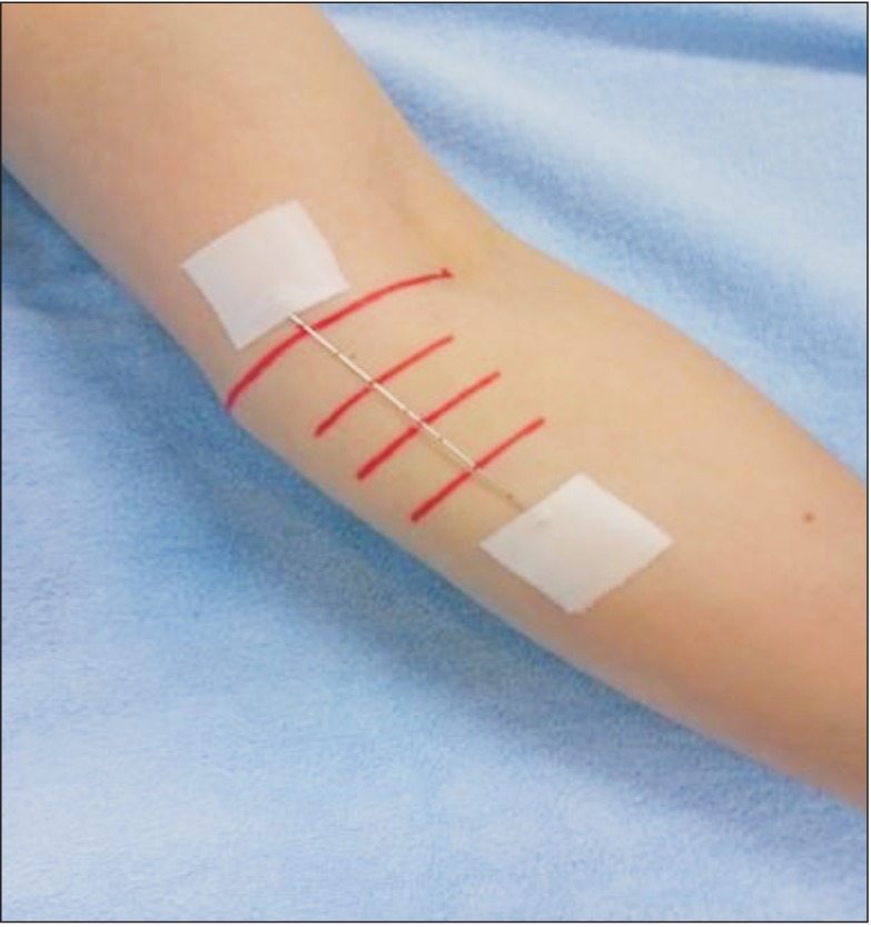
Fig. 1 Ultrasonography of the forearm with the transducer located at the four sites of the forearm in transverse plane. Point 0, the mid-point between the biceps tendon and medial epicondyle in the elbow crease; point 1, 2 cm distal to point 0; point 2, 3.5 cm distal to point 0; point 3, 5 cm distal to point 0.
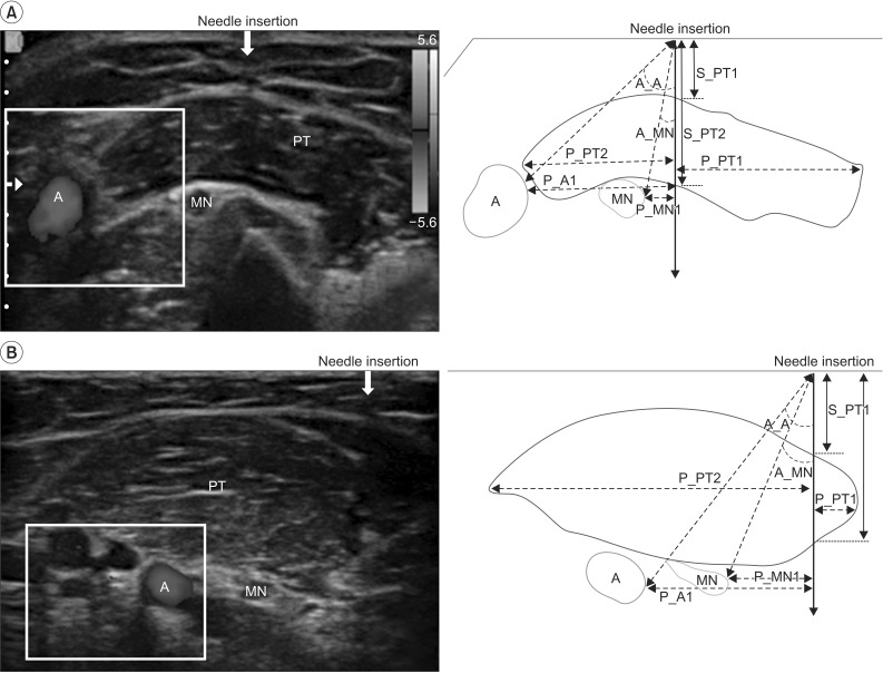
Fig. 2 Transverse view of ultrasonographic images with schematic drawing obtained at the forearm of a female at 2 cm distal to the elbow crease (point 1; A) and at 3.5 cm distal to the elbow crease (point 2; B). Needle insertion site presents as a hypoechoic focus with well-defined posterior acoustic shadowing (arrow). PT, pronator teres muscle; A, ulnar artery; MN, median nerve; S_PT1, the distance between each point and the upper margin of the pronator teres; S_PT2, the distance between each point and the lower margin of the pronator teres; P_PT1, the distance between the vertical line of each point and the medial margin of the pronator teres; P_PT2, the distance between the vertical line of each point and the lateral margin of the pronator teres; P_MN, the distance between the vertical line of each point and the medial margin median nerve; P_A, the distance between the vertical line of each point and the medial margin of brachial or ulnar artery; A_MN, the angle between the vertical line of the each point and the medial border of the median nerve; A_A, the angle between the vertical line of the each point and the medial border of the brachial or ulnar artery.
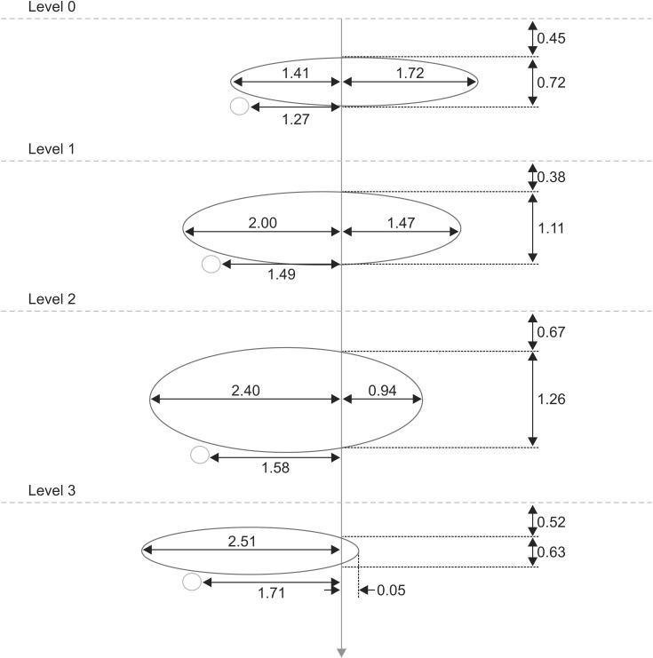
Fig. 3 In men, a schematic drawing of relationship of the distance between the vertical needle insertion line (red arrow) and the medial or lateral margin of the pronator teres (orange oval), the distance between the vertical needle insertion line and the medial margin of median nerve (yellow circle), and the distance between the skin (yellow dot line) and upper or lower margin of pronator teres.
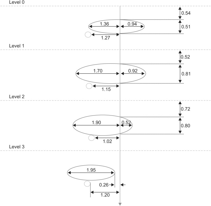
Fig. 4 In women, schematic drawing of relationship the distance between the vertical needle insertion line (red arrow) and the medial or lateral margin of the pronator teres (orange oval), the distance between the vertical needle insertion line and the medial margin of the median nerve (yellow circle), and the distance between skin (yellow dot line) and upper or lower margin of pronator teres.
Reference
-
1. Perotto AO. Anatomical guide for the electromyographer: the limb and trunk. 3rd ed. Springfield: Charles C Thomas Publisher;1994.2. Preston DC, Shapiro BE. Electromyography and neuromuscular disorders: clinical-electrophysiologic correlations. 3rd ed. New York: Elsevier Saunders;2013.3. Al-Shekhlee A, Shapiro BE, Preston DC. Iatrogenic complications and risks of nerve conduction studies and needle electromyography. Muscle Nerve. 2003; 27:517–526. PMID: 12707972.
Article4. Lee HJ, DeLisa JA. Manual of nerve conduction study and surface anatomy for needle electromyography. 4th ed. Philadelphia: Lippincott Wilkins and Williams;2005.5. Zupko RE. A dictionary of weights and measures for the British Isles: the Middle Ages to the twentieth century. Philadelphia: American Philosophical Society;1985.6. Canovas F, Mouilleron P, Bonnel F. Biometry of the muscular branches of the median nerve to the forearm. Clin Anat. 1998; 11:239–245. PMID: 9652538.
Article7. Rodriguez-Niedenfuhr M, Vazquez T, Nearn L, Ferreira B, Parkin I, Sanudo JR. Variations of the arterial pattern in the upper limb revisited: a morphological and statistical study, with a review of the literature. J Anat. 2001; 199(Pt 5):547–566. PMID: 11760886.8. Jenkins DB. Hollinshead's functional anatomy of the limbs and back. 9th ed. St. Louis: Saunders;2009.9. Lee SM, Kim K, Lee SM, Lee HS. Ultrasonographic evaluation of needle insertion site for the flexor pollicis longus. Ann Rehabil Med. 2013; 37:215–220. PMID: 23705116.
Article10. Won SJ, Kim JY, Yoon JS, Kim SJ. Ultrasonographic evaluation of needle electromyography insertion into the tibialis posterior using a posterior approach. Arch Phys Med Rehabil. 2011; 92:1921–1923. PMID: 21839985.
Article11. Rha DW, Im SH, Lee SC, Kim SK. Needle insertion into the tibialis posterior: ultrasonographic evaluation of an anterior approach. Arch Phys Med Rehabil. 2010; 91:283–287. PMID: 20159135.
Article12. Kim IJ, Kang HS, Kim BJ, Kim LN, Hwang MR, Kim DH. Comparison of the electromyography needle insertion sites for the pronator teres muscle. J Korean Assoc EMG Electrodiagn Med. 2010; 12:83–86.13. Chu-Andrews J, Johnson RJ. Electrodiagnosis: an anatomical and clinical approach. Philadelphia: Lippincott Williams & Wilkins;1986.14. Lee HJ, DeLisa JA. Surface anatomy for clinical needle electromyography. New York: Demos Medical Publishing;2000.
- Full Text Links
-
- Actions
-
Cited
- CITED
-
- Close
- Share
-
- Similar articles
-
- Early Surgical Treatment of Pronator Teres Syndrome
- Topographic Anatomy of the Median Nerve and the Pronator Teres and the Flexor Digitorum Superficialis Muscles
- Partial Tear of Pronator Teres Muscle in Amateur Golfer: A Case Report
- Abscess Formation Involving the Falciform Ligament and Ligamentum Teres
- Accuracy of Needle Placement in Cadavers: Non-Guided Versus Ultrasound-Guided
