Effect of fangchinoline on root resorption during rat orthodontic tooth movement
- Affiliations
-
- 1Department of Orthodontics, School of Stomatology, Jilin University, Changchun, China. humin@jlu.edu.cn
- KMID: 2272262
- DOI: http://doi.org/10.4041/kjod.2012.42.3.138
Abstract
OBJECTIVE
To evaluate the short-term effect of fangchinoline, an antiinflammatory drug widely used in Asia, on root resorption that is associated with orthodontic tooth movement.
METHODS
Twenty-four Wistar rats were randomly divided into 6 groups. Mesial forces of 0, 50, or 100 g were applied to the maxillary first molar of the rats in each group for 14 days by activating nickel-titanium closed-coil springs. One-half of the rats receiving each of these treatments also received injections of 200 microL fangchinoline every 2 days. Finally, movement of the maxillary first molars was measured using digitized radiographs. The molars were extracted and the surfaces of the root resorption craters were recorded using a scanning electron microscope. The distance the molars moved and resorptionarea ratio was measured, and results were analyzed using 2-way ANOVA tests.
RESULTS
There were no statistical differences in the distances the first molars moved under 50 or 100 g force, regardless of treatment with fangchinoline. However, the resorption area ratios were significantly smaller in those rats that were treated with both tension and fangchinoline than in those rats treated by tension alone.
CONCLUSIONS
Fangchinoline reduced the resorption area ratio in rats and is therefore an important means of alleviating root resorption.
Keyword
MeSH Terms
Figure
-
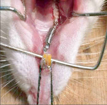
Figure 1 Rat tooth movement model. A nickel-titanium coil spring was activated after insertion between the first molar and incisors in order to move the molar mesially.
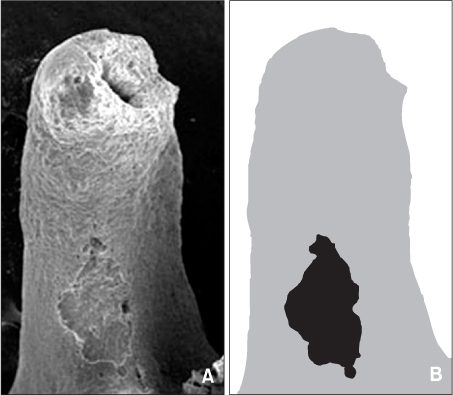
Figure 2 Calculation of the resorption area ratio in the molars of Wistar rats. A, Representative scanning electron microscope micrograph of the mesial surface of the distopalatal root. B, Diagram indicating the area of the crater (black) as compared to the total root surface area (gray). The resorption area ratio was calculated by dividing the area of the crater by the total root surface area (resorption area ratio = black area/gray area).

Figure 3 Representative scanning electron microscope micrographs of the distal root surface of molars from Wistar rats. A, 50 g force and no fangchinoline; B, 50 g force with injection of 40 µL of 5 µg/µL fangchinoline every 2 days; C, 100 g force and no fangchinoline; D, 100 g force with injection of 40 µL of 5 µg/µL fangchinoline every 2 days. Different sizes of resorption craters can be observed on the mesial surface of the root image from each group. Craters are found primarily on the cervical and middle one third of each root (white arrow).
Reference
-
1. Brudvik P, Rygh P. Non-clast cells start orthodontic root resorption in the periphery of hyalinized zones. Eur J Orthod. 1993. 15:467–480.
Article2. Segal GR, Schiffman PH, Tuncay OC. Meta analysis of the treatment-related factors of external apical root resorption. Orthod Craniofac Res. 2004. 7:71–78.
Article3. Sringkarnboriboon S, Matsumoto Y, Soma K. Root resorption related to hypofunctional periodontium in experimental tooth movement. J Dent Res. 2003. 82:486–490.
Article4. Harokopakis-Hajishengallis E. Physiologic root resorption in primary teeth: molecular and histological events. J Oral Sci. 2007. 49:1–12.
Article5. Meikle MC. The tissue, cellular, and molecular regulation of orthodontic tooth movement: 100 years after Carl Sandstedt. Eur J Orthod. 2006. 28:221–240.
Article6. Onai N, Tsunokawa Y, Suda M, Watanabe N, Nakamura K, Sugimoto Y, et al. Inhibitory effects of bisbenzylisoquinoline alkaloids on induction of proinflammatory cytokines, interleukin-1 and tumor necrosis factor-alpha. Planta Med. 1995. 61:497–501.
Article7. Choi HS, Kim HS, Min KR, Kim Y, Lim HK, Chang YK, et al. Anti-inflammatory effects of fangchinoline and tetrandrine. J Ethnopharmacol. 2000. 69:173–179.
Article8. Kim HS, Zhang YH, Oh KW, Ahn HY. Vasodilating and hypotensive effects of fangchinoline and tetrandrine on the rat aorta and the stroke-prone spontaneously hypertensive rat. J Ethnopharmacol. 1997. 58:117–123.
Article9. Zhang YH, Fang LH, Ku BS. Fangchinoline inhibits rat aortic vascular smooth muscle cell proliferation and cell cycle progression through inhibition of ERK1/2 activation and c-fos expression. Biochem Pharmacol. 2003. 66:1853–1860.
Article10. Kim HS, Zhang YH, Yun YP. Effects of tetrandrine and fangchinoline on experimental thrombosis in mice and human platelet aggregation. Planta Med. 1999. 65:135–138.
Article11. Gülçin I, Elias R, Gepdiremen A, Chea A, Topal F. Antioxidant activity of bisbenzylisoquinoline alkaloids from Stephania rotunda: cepharanthine and fangchinoline. J Enzyme Inhib Med Chem. 2010. 25:44–53.
Article12. Gonzales C, Hotokezaka H, Yoshimatsu M, Yozgatian JH, Darendeliler MA, Yoshida N. Force magnitude and duration effects on amount of tooth movement and root resorption in the rat molar. Angle Orthod. 2008. 78:502–509.
Article13. Reitan K, Kvam E. Comparative behavior of human and animal tissue during experimental tooth movement. Angle Orthod. 1971. 41:1–14.14. Storey E. The nature of tooth movement. Am J Orthod. 1973. 63:292–314.
Article15. Wang Z, McCauley LK. Osteoclasts and odontoclasts: signaling pathways to development and disease. Oral Dis. 2011. 17:129–142.
Article16. Oshiro T, Shibasaki Y, Martin TJ, Sasaki T. Immunolocalization of vacuolar-type H+-ATPase, cathepsin K, matrix metalloproteinase-9, and receptor activator of NFkappaB ligand in odontoclasts during physiological root resorption of human deciduous teeth. Anat Rec. 2001. 264:305–311.
Article17. Tsuji Y, Yamaza T, Kido MA, Goto T, Nakata S, Akamine A, et al. Expression of cathepsin K mRNA and protein in odontoclasts after experimental tooth movement in the mouse maxilla by in situ hybridization and immunoelectron microscopy. Cell Tissue Res. 2001. 303:359–369.
Article18. Zhuang L, Meng X, Li P, Bai Y. A pilot study on three dimensional morphology of root by Micro-CT during orthodontic root resorption. J Modern Stomatol. 2010. 24:35–38.19. Inaoka T, Bilbe G, Ishibashi O, Tezuka K, Kumegawa M, Kokubo T. Molecular cloning of human cDNA for cathepsin K: novel cysteine proteinase predominantly expressed in bone. Biochem Biophys Res Commun. 1995. 206:89–96.
Article20. Henriksen K, Tanko LB, Qvist P, Delmas PD, Christiansen C, Karsdal MA. Assessment of osteoclast number and function: application in the development of new and improved treatment modalities for bone diseases. Osteoporos Int. 2007. 18:681–685.
Article21. Jäger A, Zhang D, Kawarizadeh A, Tolba R, Braumann B, Lossdörfer S, et al. Soluble cytokine receptor treatment in experimental orthodontic tooth movement in the rat. Eur J Orthod. 2005. 27:1–11.
Article
- Full Text Links
-
- Actions
-
Cited
- CITED
-
- Close
- Share
-
- Similar articles
-
- Changes in the titer of tooth root antibodies accompanying root resorption associated with orthodontic tooth movement
- A study on the affecting factors on root resorption
- Effects of continuous force application for extrusive tipping movement on periapical root resorption in the rat mandibular first molar
- Effect of orthodontic force on the amount of tooth movement and root resorption in rat
- Mechanisms of Osteoclastogenesis in Orthodontic Tooth Movement and Orthodontically Induced Tooth Root Resorption
