Effects of the Off-Loading Brace on the Activation of Femoral Muscles: A Preliminary Study
- Affiliations
-
- 1Department of Rehabilitation Medicine, Hallym University College of Medicine, Chuncheon 200-704, Korea. newbabylon@naver.com
- KMID: 2266816
- DOI: http://doi.org/10.5535/arm.2011.35.6.887
Abstract
OBJECTIVE
To provide the off-loading knee brace was designed relief for the pain associated with osteoarthritis by reduce loads on the degenerative compartment of the knee. This study examined the effects of the off-loading knee brace on activation of femoral muscles during squatting, slow and fast walking exercise in healthy young individuals. METHOD: Ten healthy male subjects without a history of knee pain were recruited. Each subject was asked to do squatting, slow and fast walking exercises with a brace secured to the dominant leg. The same exercises were repeated without the brace. Surface electromyographic (sEMG) data was collected from the vastus medialis oblique (VMO), vastus lateralis (VL) and biceps femoris (BF) muscles from the dominant side of the leg. All dynamic root mean squre (RMS) values of sEMG were standardized to static RMS values of the maximal isometric contraction and expressed as a percentage of maximal activity.
RESULTS
We found that VMO activity was significantly decreased with application of the off-loading knee brace during squatting and fast walking exercise. However there were no significant differences in VMO activity with application of the off-loading knee brace during slow walking exercise.
CONCLUSION
These results suggest that the external moment of the brace which effectively stabilized the patella in the movement in which the knee joints become relatively unstable. The brace could be useful in the short term, but for long-term use, weakening of the VMO is predicted. Therefore the program of selective muscular strength strengthening for the VMO should be emphasized.
Keyword
MeSH Terms
Figure
-
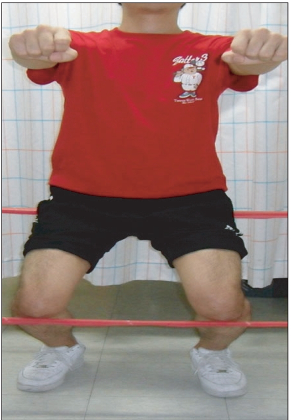
Fig. 1 Performing the squatting exercise with 90° of knee flexion using fixed bars.
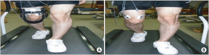
Fig. 2 Demonstration of subjects as they performed walking exercise with brace to the dominant leg (A), and without brace (B).
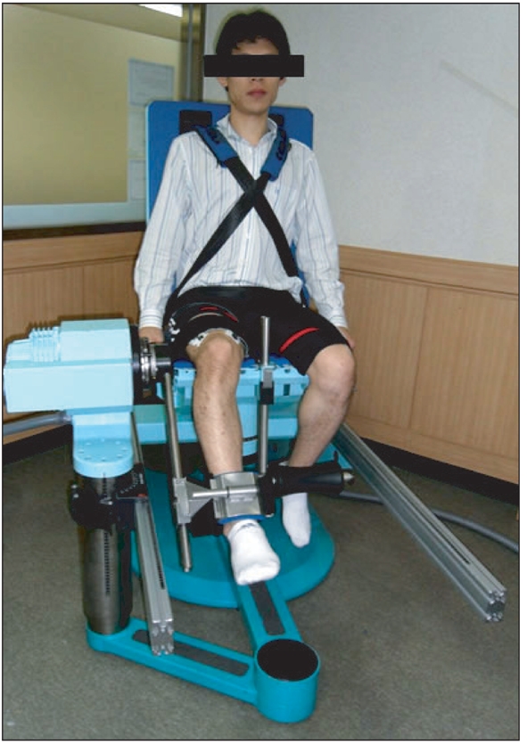
Fig. 3 Measurement of maximal isometric voluntary contraction at 45° of knee flexion on an isokinetic dynamometer with surface electriomyographic activities of the VMO, VL and BF muscles. VMO: Vastus medialis oblique, VL: Vastus lateralis, BF: Biceps femoris.
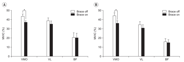
Fig. 4 The changes of electromyographic activities expressed as percentage of maximal voluntary isometric contraction of the VMO, VL and BF muscles during squatting exercise. (A) extension phase, (B) flexion phase. VMO: Vastus medialis oblique, VL: Vastus lateralis, BF: Biceps femoris, MVIC (%): Percentage of maximal voluntary isometric contraction; *p<0.05.
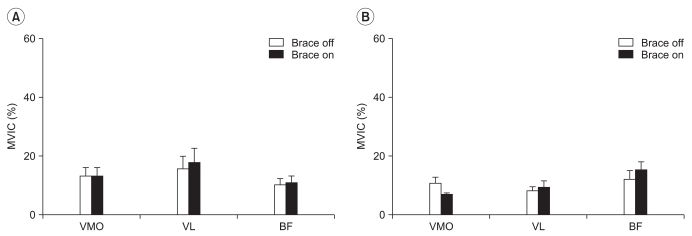
Fig. 5 The changes of electromyographic activities (expressed as percentage of maximal voluntary isometric contraction) of VMO, VL and BF muscles during slow walking exercise. (A) stance phase, (B) swing phase. VMO: Vastus medialis obique VL: Vastus lateralis BF: Biceps femoris, MVIC (%): percentage of maximal voluntary isometric contraction; *p<0.05.
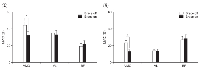
Fig. 6 The changes of electromyographic activities expressed as percentage of maximal voluntary isometric contraction of VMO, VL and BF muscles during fast walking exercise. (A) stance phase, (B) swing phase. VMO: Vastus medialis obique VL: Vastus lateralis BF: Biceps femoris, MVIC (%): percentage of maximal voluntary isometric contraction; *p<0.05.
Reference
-
1. Cho HJ, Chang CB, Kim KW, Park JH, Yoo JH, Koh IJ, Kim TK. Gender and prevalence of knee osteoarthritis types in elderly Koreans. J Arthroplasty. 2011; 26:994–999. PMID: 21414750.
Article2. Kim I, Kim HA, Seo YI, Song YW, Jeong JY, Kim DH. The prevalence of knee osteoarthritis in elderly community residents in Korea. J Korean Med Sci. 2010; 25:293–298. PMID: 20119586.
Article3. Hurley MV. Muscle dysfunction and effective rehabilitation of knee osteoarthritis: what we know and what we need to find out. Arthritis Rheum. 2003; 49:444–452. PMID: 12794802.
Article4. Pollo FE, Jackson RW. Knee bracing for unicompartmental osteoarthritis. J Am Acad Orthop Surg. 2006; 14:5–11. PMID: 16394162.
Article5. Rannou F, Poiraudeau S, Beaudreuil J. Role of bracing in the management of knee osteoarthritis. Curr Opin Rheumatol. 2010; 22:218–222. PMID: 20035222.
Article6. Ahlback S. Osteoarthrosis of the knee. A radiographic investigation. Acta Radiol Diagn (Stockh). 1968; (Suppl 277):7–72. PMID: 5706059.7. Felson DT, Radin EL. What causes knee osteoarthrosis: are different compartments susceptible to different risk factors? J Rheumatol. 1994; 21:181–183. PMID: 8182619.8. Marks R, Penton L. Are foot orthotics efficacious for treating painful medial compartment knee osteoarthritis? A review of the literature. Int J Clin Pract. 2004; 58:49–45. PMID: 14994971.
Article9. Pham T, Maillefert JF, Hudry C, Kieffert P, Bourgeois P, Lechevalier D, Dougados M. Laterally elevated wedged insoles in the treatment of medial knee osteoarthritis. A two-year prospective randomized controlled study. Osteoarthritis Cartilage. 2004; 12:46–45. PMID: 14697682.10. Baker K, Goggins J, Xie H, Szumowski K, LaValley M, Hunter DJ, Felson DT. A randomized crossover trial of a wedged insole for treatment of knee osteoarthritis. Arthritis Rheum. 2007; 56:1198–1203. PMID: 17393448.
Article11. Hinman RS, Bowles KA, Bennell KL. Laterally wedged insoles in knee osteoarthritis: do biomechanical effects decline after one month of wear? BMC Musculoskelet Disord. 2009; 10:146. PMID: 19939281.
Article12. Dennis DA, Komistek RD, Nadaud MC, Mahfouz M. Evaluation of off-loading braces for treatment of unicompartmental knee arthrosis. J Arthroplasty. 2006; 21:2–8. PMID: 16781418.
Article13. Kirkley A, Webster-Bogaert S, Litchfield R, Amendola A, MacDonald S, McCalden R, Fowler P. The effect of bracing on varus gonarthrosis. J Bone Joint Surg Am. 1999; 81:539–548. PMID: 10225800.
Article14. Matsuno H, Kadowaki KM, Tsuji H. Generation II knee bracing for severe medial compartment osteoarthritis of the knee. Arch Phys Med Rehabil. 1997; 78:745–749. PMID: 9228878.
Article15. Pollo FE, Otis JC, Backus SI, Warren RF, Wickiewicz TL. Reduction of medial compartment loads with valgus bracing of the osteoarthritic knee. Am J Sports Med. 2002; 30:414–421. PMID: 12016084.
Article16. Schmalz T, Knopf E, Drewitz H, Blumentritt S. Analysis of biomechanical effectiveness of valgus-inducing knee brace for osteoarthritis of knee. J Rehabil Res Dev. 2010; 47:419–429. PMID: 20803386.
Article17. Hebbal GV, Mysorekar VR. Evaluation of some tasks used for specifying handedness and footedness. Percept Mot Skills. 2006; 102:163–164. PMID: 16671615.
Article18. Cram JR, Kasman GS, Holtz J. Introduction to surface electromyography. 1998. 1st ed. Maryland: Aspen;p. 363–368.19. Winter DA, Robertson DG. Joint torque and energy patterns in normal gait. Biol Cybern. 1978; 29:137–142. PMID: 667202.20. Harrington IJ. Static and dynamic loading patterns in knee joints with deformities. J Bone Joint Surg Am. 1983; 65:247–259. PMID: 6822587.
Article21. Johnson F, Leitl S, Waugh W. The distribution of load across the knee. A comparison of static and dynamic measurements. J Bone Joint Surg Br. 1980; 62:346–349. PMID: 7410467.
Article22. Hurley MV. The role of muscle weakness in the pathogenesis of osteoarthritis. Rheum Dis Clin North Am. 1999; 25:283–298. PMID: 10356418.
Article23. Slemenda C, Brandt KD, Heilman DK, Mazzuca S, Braunstein EM, Katz BP, Wolinsky FD. Quadriceps weakness and osteoarthritis of the knee. Ann Intern Med. 1997; 127:97–104. PMID: 9230035.
Article24. Collado H, Fredericson M. Patellofemoral pain syndrome. Clin Sports Med. 2010; 29:379–398. PMID: 20610028.
Article25. Toumi H, Poumarat G, Benjamin M, Best TM, F'Guyer S, Fairclough J. New insights into the function of the vastus medialis with clinical implications. Med Sci Sports Exerc. 2007; 39:1153–1159. PMID: 17596784.
Article26. Perry J, Burnfield JM. Gait analysis: normal and pathological function. 2010. 2nd ed. New Jersey: SLACK;p. 385–517.27. Perry J, Burnfield JM. Gait analysis: normal and pathological function. 2010. 2nd ed. New Jersey: SLACK;p. 88–100.
- Full Text Links
-
- Actions
-
Cited
- CITED
-
- Close
- Share
-
- Similar articles
-
- The Treatment of Femoral Shaft Fractures by Cast Brace
- Clinical Study on Cast-Brace in Femoral Fractures
- Relapse of Clubfoot after Treatment with the Ponseti Method and the Function of the Foot Abduction Orthosis
- A Rotation - plasty for Focal Femoral Deficiency Due to Chronic Osteomyelitis
- A novel finger brace for preventing finger stiffness after trauma or surgery: a preliminary report with a case series
