Ann Rehabil Med.
2013 Dec;37(6):896-900. 10.5535/arm.2013.37.6.896.
Thoracic Outlet Syndrome Caused by Schwannoma of Brachial Plexus
- Affiliations
-
- 1Department of Physical Medicine and Rehabilitation, Kyung Hee University School of Medicine, Seoul, Korea. id-login@hanmail.net
- KMID: 2266571
- DOI: http://doi.org/10.5535/arm.2013.37.6.896
Abstract
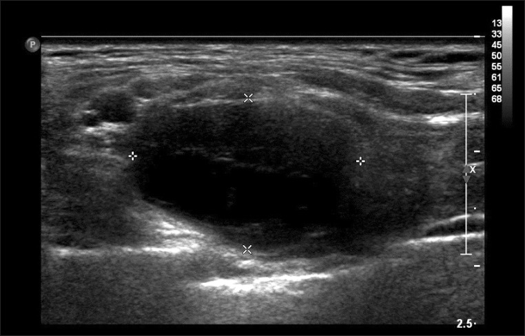
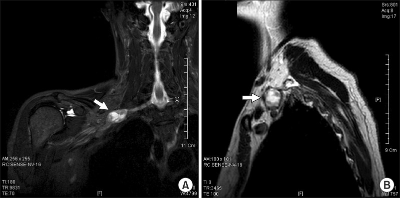
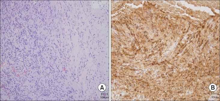
- Schwannomas are benign, usually slow-growing tumors that originate from Schwann cells surrounding peripheral, cranial, or autonomic nerves. The most common form of these tumors is acoustic neuroma. Schwannomas of the brachial plexus are quite rare, and symptomatic schwannomas of the brachial plexus are even rarer. A 47-year-old woman presented with a 1-year history of dysesthesia, neuropathic pain, and mild weakness of the right upper limb. Results of physical examination and electrodiagnostic studies supported a diagnosis as thoracic outlet syndrome. Conservative treatment did not relieve her symptoms. After 9 months, a soft mass was found at the upper margin of the right clavicle. Magnetic resonance imaging showed a 3.0x1.8x1.7 cm ovoid mass between the inferior trunk and the anterior division of the brachial plexus. Surgical mass excision and biopsy were performed. Pathological findings revealed the presence of schwannoma. After schwannoma removal, the right hand weakness did not progress any further and neuropathic pain gradually reduced. However, dysesthesia at the right C8 and T1 dermatome did not improve.
MeSH Terms
Figure
Reference
-
1. Peet RM, Henriksen JD, Anderson TP, Martin GM. Thoracic-outlet syndrome: evaluation of a therapeutic exercise program. Proc Staff Meet Mayo Clin. 1956; 31:281–287. PMID: 13323047.2. Sanders RJ, Hammond SL, Rao NM. Thoracic outlet syndrome: a review. Neurologist. 2008; 14:365–373. PMID: 19008742.3. Kumar A, Akhtar S. Schwannoma of brachial plexus. Indian J Surg. 2011; 73:80–81. PMID: 22211049.
Article4. Chen F, Miyahara R, Matsunaga Y, Koyama T. Schwannoma of the brachial plexus presenting as an enlarging cystic mass: report of a case. Ann Thorac Cardiovasc Surg. 2008; 14:311–313. PMID: 18989247.5. Nichols AW. Diagnosis and management of thoracic outlet syndrome. Curr Sports Med Rep. 2009; 8:240–249. PMID: 19741351.
Article6. Crosby CA, Wehbe MA. Conservative treatment for thoracic outlet syndrome. Hand Clin. 2004; 20:43–49. PMID: 15005383.
Article7. Novak CB. Conservative management of thoracic outlet syndrome. Semin Thorac Cardiovasc Surg. 1996; 8:201–207. PMID: 8672574.8. Kim DS. Neurogenic tumor of the brachial plexus: a case report. Korean J Thorac Cardiovasc Surg. 2004; 37:84–87.9. Kim YW, Ahn SK, Song JH. A case of brachial plexus schwannoma. J Korean Neurosurg Soc. 2006; 39:396–399.10. Cho DG, Son BC, Cho KD, Jo MS, Wang YP. Microsurgical resection of schwannoma of the brachial plexus: a case report. Korean J Thorac Cardiovasc Surg. 2005; 38:249–252.
- Full Text Links
- Actions
-
Cited
- CITED
-
- Close
- Share
- Similar articles
-
- Progressive Brachial Plexus Palsy after Fixation of Clavicle Shaft Nonunion: A Case Report
- Thoracic Outlet Syndrome Induced by Huge Lipoma: A Case Report
- Transaxillary Approach for First Rib Resection to Relieve Thoracic Outlet Syndrome
- A Surgical Resection of Giant Schwannoma in the Brachial lexus
- En Bloc Resection of a Thoracic Outlet for a Recurred Malignant Schwannoma of the Brachial Plexus: A case report


