Ultrasound-Guided Myofascial Trigger Point Injection Into Brachialis Muscle for Rotator Cuff Disease Patients With Upper Arm Pain: A Pilot Study
- Affiliations
-
- 1Department of Rehabilitation Medicine and Research Institute of Rehabilitation Medicine, Yonsei University College of Medicine, Seoul, Korea. bettertomo@yuhs.ac
- 2Department of Physical Medicine and Rehabilitation, Samsung Medical Center, Sungkyunkwan University School of Medicine, Seoul, Korea.
- KMID: 2266502
- DOI: http://doi.org/10.5535/arm.2014.38.5.673
Abstract
OBJECTIVE
To assess the efficacy of trigger point injection into brachialis muscle for rotator cuff disease patients with upper arm pain.
METHODS
A prospective, randomized, and single-blinded clinical pilot trial was performed at university rehabilitation hospital. Twenty-one patients clinically diagnosed with rotator cuff disease suspected of having brachialis myofascial pain syndrome (MPS) were randomly allocated into two groups. Effect of ultrasound (US)-guided trigger point injection (n=11) and oral non-steroidal anti-inflammatory drug (NSAID) (n=10) was compared by visual analog scale (VAS).
RESULTS
US-guided trigger point injection of brachialis muscle resulted in excellent outcome compared to the oral NSAID group. Mean VAS scores decreased significantly after 2 weeks of treatment compared to the baseline in both groups (7.3 vs. 4.5 in the injection group and 7.4 vs. 5.9 in the oral group). The decrease of the VAS score caused by injection (capital DE, CyrillicVAS=-2.8) was significantly larger than caused by oral NSAID (capital DE, CyrillicVAS=-1.5) (p<0.05).
CONCLUSION
In patients with rotator cuff disease, US-guided trigger point injection of the brachialis muscle is safe and effective for both diagnosis and treatment when the cause of pain is suspected to be originated from the muscle.
MeSH Terms
Figure
-
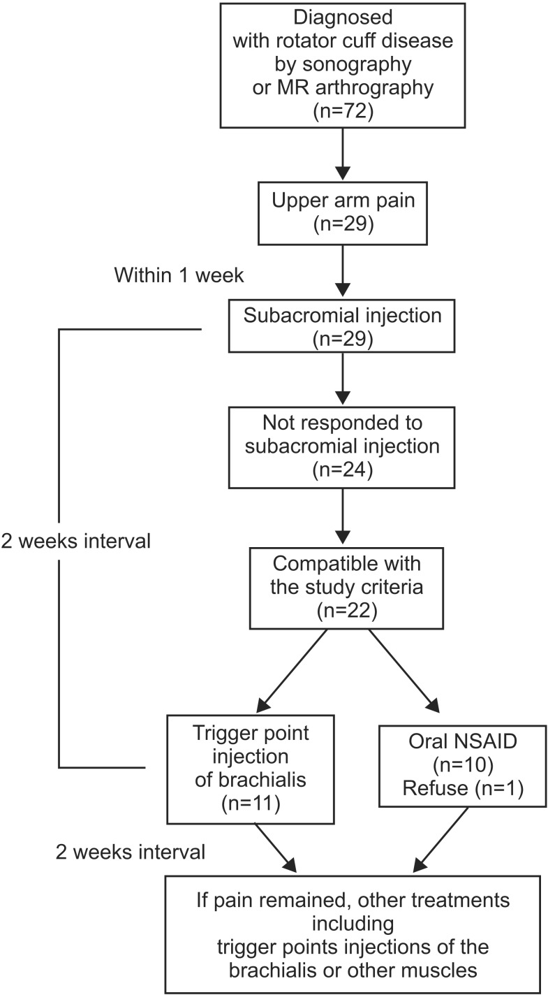
Fig. 1 Design of the study. MR, magnetic resonance; NSAID, non-steroidal anti-inflammatory drug.
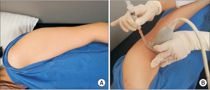
Fig. 2 Proper positioning during injection procedure. (A) The patient is laid on the lateral side with the affected side up, with the upper arm placed tightly to the trunk to minimize the motion during injection. (B) The probe is positioned on the maximal tender point to show the transverse image of the brachialis muscle, and the needle is approached with inplane method.
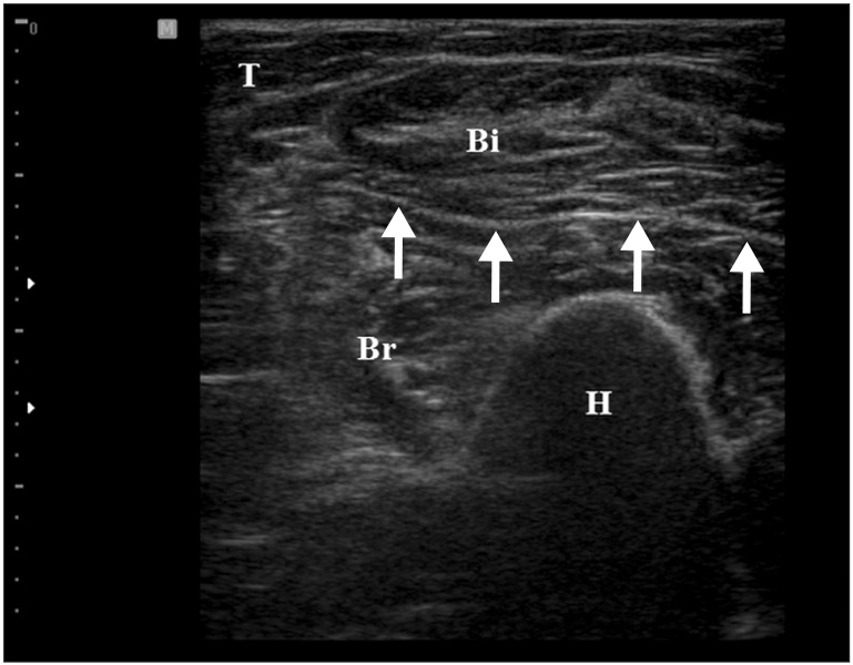
Fig. 3 Transverse ultrasound image of the brachialis muscle. The brachialis muscle (Br) is seen below the biceps brachii muscle (Bi). H, humerus; T, triceps.
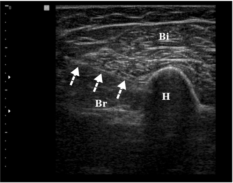
Fig. 4 Transverse ultrasound image by the in-plane method. Needle passage into the target muscle (Br) is visualized (arrow). Bi, biceps brachii; Br, brachialis; H, humerus.
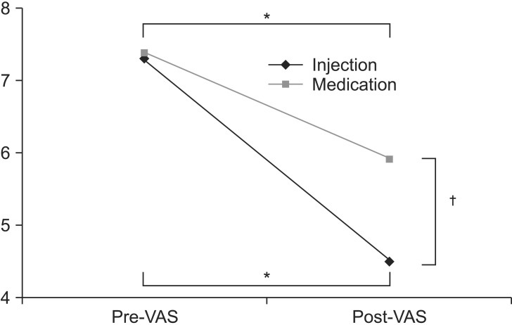
Fig. 5 Effect of each treatment. VAS changes significantly after injection and medication (*p<0.05) and VAS in injection group decreases more compared to the medication group after treatment (†p<0.05). VAS, visual analogue scale; pre-VAS, VAS at pretreatment; post-VAS, VAS after 2 weeks of treatment.
Cited by 1 articles
-
Characteristics of Myofascial Pain Syndrome of the Infraspinatus Muscle
Junbeom Kwon, Hyoung Seop Kim, Won Hyuk Chang, Chunung Park, Sang Chul Lee
Ann Rehabil Med. 2017;41(4):573-581. doi: 10.5535/arm.2017.41.4.573.
Reference
-
1. Toivonen DA, Tuite MJ, Orwin JF. Acromial structure and tears of the rotator cuff. J Shoulder Elbow Surg. 1995; 4:376–383. PMID: 8548441.
Article2. Andrews JR. Diagnosis and treatment of chronic painful shoulder: review of nonsurgical interventions. Arthroscopy. 2005; 21:333–347. PMID: 15756189.
Article3. Chou WY, Ko JY, Wang FS, Huang CC, Wong T, Wang CJ, et al. Effect of sodium hyaluronate treatment on rotator cuff lesions without complete tears: a randomized, double-blind, placebo-controlled study. J Shoulder Elbow Surg. 2010; 19:557–563. PMID: 19963403.
Article4. Hong CZ. Pathophysiology of myofascial trigger point. J Formos Med Assoc. 1996; 95:93–104. PMID: 9064014.5. Hong CZ, Simons DG. Pathophysiologic and electrophysiologic mechanisms of myofascial trigger points. Arch Phys Med Rehabil. 1998; 79:863–872. PMID: 9685106.
Article6. Hong CZ. Myofascial pain therapy. J Musculoskelet Pain. 2004; 12:37–43.
Article7. Bron C, Wensing M, Franssen JL, Oostendorp RA. Treatment of myofascial trigger points in common shoulder disorders by physical therapy: a randomized controlled trial (ISRCTN75722066). BMC Musculoskelet Disord. 2007; 8:107. PMID: 17983467.
Article8. Bron C, Dommerholt J, Stegenga B, Wensing M, Oostendorp RA. High prevalence of shoulder girdle muscles with myofascial trigger points in patients with shoulder pain. BMC Musculoskelet Disord. 2011; 12:139. PMID: 21711512.
Article9. Calais-Germain B. The elbow. Anatomy of movement. Seatle: Eastland Press;1993. p. 131–146.10. Hong CZ. Specific sequential myofascial trigger point therapy in the treatment of a patient with myofascial pain syndrome associated with reflex sympathetic dystrophy. Australas Chiropr Osteopathy. 2000; 9:7–11. PMID: 17987165.11. Travell JG, Simons DG. Myofascial pain and dysfunction: the trigger point manual. 2nd ed. Baltimore: Williams & Wilkins;1999.12. Rha DW, Shin JC, Kim YK, Jung JH, Kim YU, Lee SC. Detecting local twitch responses of myofascial trigger points in the lower-back muscles using ultrasonography. Arch Phys Med Rehabil. 2011; 92:1576–1580.e1. PMID: 21839982.
Article13. Kim DS, Jeong TY, Kim YK, Chang WH, Yoon JG, Lee SC. Usefulness of a myofascial trigger point injection for groin pain in patients with chronic prostatitis/ chronic pelvic pain syndrome: a pilot study. Arch Phys Med Rehabil. 2013; 94:930–936. PMID: 23262156.14. Muscolino JE. The muscle and bone palpation manual with trigger points, referral patterns, and stretching. 1st ed. St. Louis: Mosby/Elsevier;2009.15. Landin D, Myers J, Thompson M, Castle R, Porter J. The role of the biceps brachii in shoulder elevation. J Electromyogr Kinesiol. 2008; 18:270–275. PMID: 17196396.
Article16. Pagnani MJ, Deng XH, Warren RF, Torzilli PA, O'Brien SJ. Role of the long head of the biceps brachii in glenohumeral stability: a biomechanical study in cadavera. J Shoulder Elbow Surg. 1996; 5:255–262. PMID: 8872922.
Article17. Leonello DT, Galley IJ, Bain GI, Carter CD. Brachialis muscle anatomy: a study in cadavers. J Bone Joint Surg Am. 2007; 89:1293–1297. PMID: 17545433.18. Rispoli DM, Athwal GS, Sperling JW, Cofield RH. The anatomy of the deltoid insertion. J Shoulder Elbow Surg. 2009; 18:386–390. PMID: 19186076.
Article19. McCully SP, Suprak DN, Kosek P, Karduna AR. Suprascapular nerve block results in a compensatory increase in deltoid muscle activity. J Biomech. 2007; 40:1839–1846. PMID: 17034796.
Article20. DeLisa JA, Gans BM, Bockenek WL, Frontera WR, Gerber LH, Geiringer SR, et al. Physical medicine and rehabilitation: principles and practice. 4th ed. Philadelphia: Lippincott Williams & Wilkins;2005.21. Stevens KJ, Crain JM, Akizuki KH, Beaulieu CF. Imaging and ultrasound-guided steroid injection of internal oblique muscle strains in baseball pitchers. Am J Sports Med. 2010; 38:581–585. PMID: 20051499.
Article
- Full Text Links
-
- Actions
-
Cited
- CITED
-
- Close
- Share
-
- Similar articles
-
- Treatment Experience of Pulsed Radiofrequency Under Ultrasound Guided to the Trapezius Muscle at Myofascial Pain Syndrome: A Case Report
- Ultrasound Guided Thoracic Paravertebral Space Block for Chronic Intractable Upper Back Pain
- Trigger Point Injection for the Treatment of Myofascial Pain Syndrome
- Application of Ultrasound-Guided Trigger Point Injection for Myofascial Trigger Points in the Subscapularis and Pectoralis Muscles to Post-Mastectomy Patients: A Pilot Study
- Ischemic Compression After Trigger Point Injection Affect the Treatment of Myofascial Trigger Points
