Spectrophotometric Measurement of Minimal Erythema Dose Sites after Narrowband Ultraviolet B Phototesting: Clinical Implication of Spetrophotometric Values in Phototherapy
- Affiliations
-
- 1Department of Dermatology, Dong-A University College of Medicine, Busan, Korea. khkim@dau.ac.kr
- 2Department of Dermatology, Sorokdo National Hospital, Goheung, Korea.
- KMID: 2265693
- DOI: http://doi.org/10.5021/ad.2014.26.1.17
Abstract
- BACKGROUND
The spectrophotometer is well known to be a useful tool for estimating the objective minimal erythema dose (MED) during planning of phototherapy protocol. However, only a few spectrophotometric values are used to evaluate the erythema and pigmentation of the MED site during phototesting.
OBJECTIVE
To determinea new meaning of the relationships among spectrophotometric values during phototesting.
METHODS
Twenty-five patients with psoriasis and 23 patients with vitiligo were selected before undergoing narrowband ultraviolet B phototherapy. We interpreted the gross findings of erythema and measured the L*a*b* values using a spectrophotometer at each phototest spot. We compared MEDs, basic spectrophotometric values (L*a*b*), and b*/L* values separately according to skin type, and determined the correlation of each spectrophotometric value and the correlation between a* and b*/L* values.
RESULTS
Among L*a*b* values, only b* values showed a statistically significant difference between the type III and IV groups (p=0.003). There was a positive correlation only between MEDs and b* values (p<0.05). The average b*/L*value in the type IV group was significantly higher than the type III group (p<0.05).
CONCLUSION
The higher b* values in type IV skin indicates that skin tanning develops more prominently than type III. The correlation between MEDs and b* values may signify that the skin pigmentation status is deepened with the higher MEDs. The difference in b*/L*values between type III and IV skin reflects that the b*/L*value is thought to be an index of tanning. The a* value, known as an index of erythema, does not influence the degree of tanning.
Keyword
MeSH Terms
Figure
-
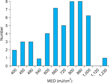
Fig. 1 The distribution of minimal erythema dose (MED)-narrowband ultraviolet B (NBUVB) in all the patients.
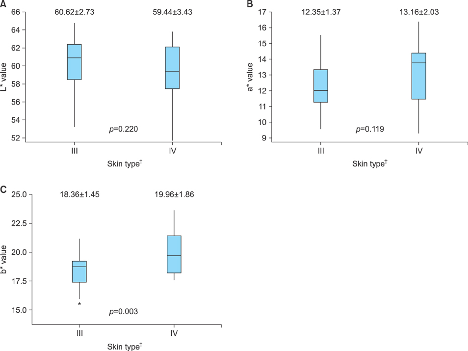
Fig. 2 The average spectrophotometric values measured at minimal erythema dose sites in skin type III and skin type IV (A: L* value; B: a* value; C: b* value). Only b* value shows statistically significant difference between the type III and IV groups (p=0.003). †Classification according to the standard of Fitzpatrick skin type III, IV.
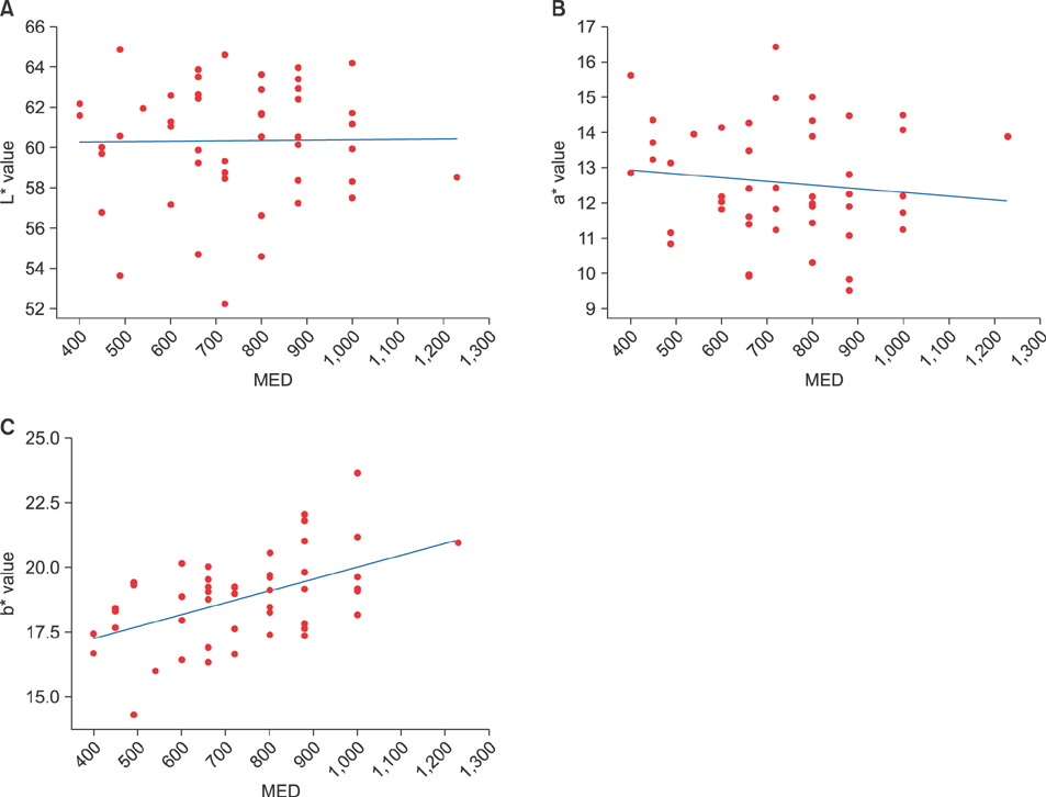
Fig. 3 A correlation between each L*, a*, b*, and minimal erythema dose (MED) values. (A) L* value=60.30±2.95, range 52.16~64.89. (B) a* value=12.57±1.60, range 9.53~16.42. (C) b* value=18.80±1.70, range, 14.31~23.64. Only correlation between b* and MED values show significant correlation (p<0.05).

Fig. 4 Significant correlation in a*L* curve (A, p<0.05). Although the b*L* curve and the b*a* curve seem to show significant correlation, but no significant correlation is shown in the b*a* curve or b*L* curve (B and C, p<0.05, respectively). †Classification according to the standard of Fitzpatrick skin type III, IV.
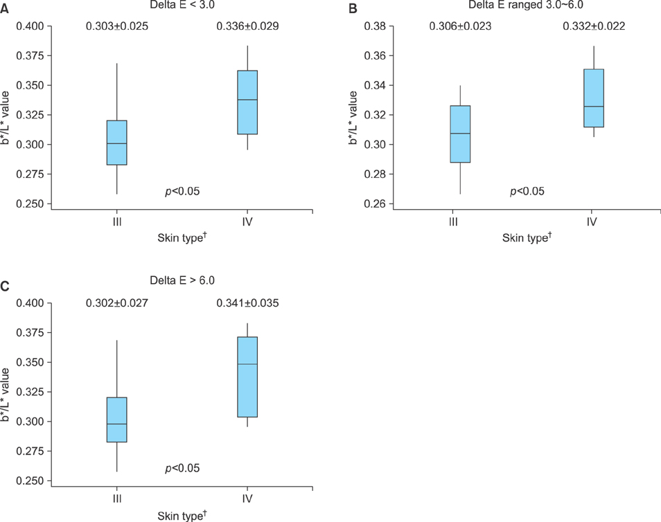
Fig. 5 The differences of b*/L* values between skin types III and IV depending on the ranges of delta E (A: <3.0; B: 3.0~6.0: C, >6.0). All data show statistically significant difference (p<0.05). †Classification according to the standard of Fitzpatrick skin type III, IV.
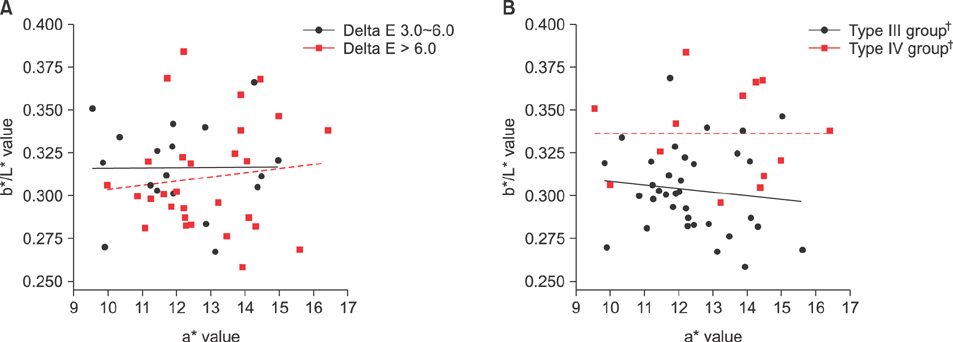
Fig. 6 A correlation between a* and b*/L* depending on (A) the delta E value and (B) in skin types. Correlation between a* values and b*/L* in skin type III and IV showed no significant correlation. †Classification according to the standard of Fitzpatrick skin type III, IV.
Reference
-
1. Rim JH, Choe YB, Youn JI. Minimal erythema dose of narrow band UVB in Korean psoriasis patients. Korean J Dermatol. 2001; 39:883–886.2. Choe YB, Park SB, Yoon JI. Narrow-band UVB phototherapy in Korean psoriasis patients. Korean J Dermatol. 2000; 38:358–362.3. Carretero-Mangolis C, Lim HW. Correlation between skin types and minimal erythema dose in narrowband UVB (TL-01) phototherapy. Photodermatol Photoimmunol Photomed. 2001; 17:244–246.
Article4. Rosbertson AR. The CIE 1976 color-difference formulae. Color Res Appl. 1977; 2:7–11.
Article5. Choi KW, Kim KH, Kim YH. Comparative study of the gross interpretation of phototesting and objective measurement with using a spectrophotometer for patients with psoriasis and vitiligo treated with narrow-band UVB. Ann Dermatol. 2009; 21:136–141.
Article6. Youn JI, Park JY, Jo SJ, Rim JH, Choe YB. Assessment of the usefulness of skin phototype and skin color as the parameter of cutaneous narrow band UVB sensitivity in psoriasis patients. Photodermatol Photoimmunol Photomed. 2003; 19:261–264.
Article7. Westerhof W, Estevez-Uscanga O, Meens J, Kammeyer A, Durocq M, Cario I. The relation between constitutional skin color and photosensitivity estimated from UV-induced erythema and pigmentation dose-response curves. J Invest Dermatol. 1990; 94:812–816.
Article8. Andreassi L, Simoni S, Fiorini P, Fimiani M. Phenotypic characters related to skin type and minimal erythemal dose. Photodermatol. 1987; 4:43–46.9. Seitz JC, Whitmore CG. Measurement of erythema and tanning responses in human skin using a tri-stimulus colorimeter. Dermatologica. 1988; 177:70–75.
Article10. Clarys P, Alewaeters K, Lambrecht R, Barel AO. Skin color measurements: comparison between three instruments: the Chromameter(R), the DermaSpectrometer(R) and the Mexameter( R). Skin Res Technol. 2000; 6:230–238.
Article11. Westerhof W, van Hasselt BA, Kammeijer A. Quantification of UV-induced erythema with a portable computer controlled chromameter. Photodermatol. 1986; 3:310–314.12. Stamatas GN, Zmudzka BZ, Kollias N, Beer JZ. Non-invasive measurements of skin pigmentation in situ. Pigment Cell Res. 2004; 17:618–626.
Article
- Full Text Links
-
- Actions
-
Cited
- CITED
-
- Close
- Share
-
- Similar articles
-
- Comparative Study of the Gross Interpretation of Phototesting and Objective Measurement with Using a Spectrophotometer for Patients with Psoriasis and Vitiligo Treated with Narrow-band UVB
- Minimal Erythema Dose of Narrow Band UVB in Korean Psoriasis Patients
- A Case of Generalized Pigmented Purpuric Dermatosis Treated with Narrowband Ultraviolet B Phototherapy in a Child
- Narrowband Ultraviolet B Phototherapy of Early Stage Mycosis Fungoides in Korean Patients
- Three Cases of Acquired Reactive Perforating Collagenosis Improved by Narrowband Ultraviolet B Phototherapy
