Ann Dermatol.
2014 Aug;26(4):551-552. 10.5021/ad.2014.26.4.551.
Pacinian Neuromas Presenting as Soft Tumors on the Volar Aspect of the Fingertips
- Affiliations
-
- 1Department of Dermatology, Eulji University School of Medicine, Daejeon, Korea. jke0224@eulji.ac.kr
- 2Department of Pathology, Eulji University School of Medicine, Daejeon, Korea.
- KMID: 2265612
- DOI: http://doi.org/10.5021/ad.2014.26.4.551
Abstract
- No abstract available.
MeSH Terms
Figure
-
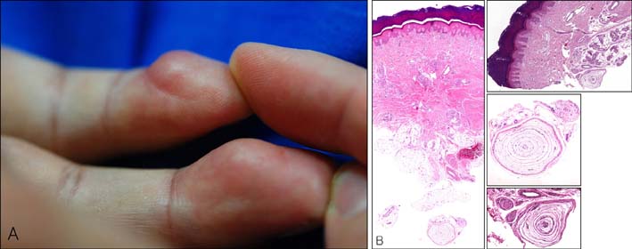
Fig. 1 (A) Flesh-colored, protruding, 1.5-cm soft tumor-like masses on the volar aspect of the right fourth and fifth fingertips. (B) Multiple (left) or enlarged (top right) Pacinian corpuscles in the subcutaneous tissue surrounded by numerous nerve fibers. Dermal fibrosis and increased adnexal tissues are also observed (H&E, ×40; small boxes: H&E, ×200).
Reference
-
1. Reznik M, Thiry A, Fridman V. Painful hyperplasia and hypertrophy of pacinian corpuscles in the hand: report of two cases with immunohistochemical and ultrastructural studies, and a review of the literature. Am J Dermatopathol. 1998; 20:203–207.2. Kenmochi A, Satoh T, Fukuyama K, Yokozeki H. Pacinian neuroma. J Eur Acad Dermatol Venereol. 2006; 20:1384–1385.
Article3. Rhode CM, Jennings WD Jr. Pacinian corpuscle neuroma of digital nerves. South Med J. 1975; 68:86–89.
Article4. Choi Y, Lim WS, Jin SY, Lee JH, Lee AY, Lee SH. Pacinian neuroma on the tips of fingers. Korean J Dermatol. 2011; 49:847–849.5. Lang-Stevenson AI. Induction of hyperplasia and hypertrophy of pacinian corpuscles. Br Med J (Clin Res Ed). 1984; 288:972–973.
Article
