A Case of Lymphomatoid Keratosis
- Affiliations
-
- 1Department of Dermatology, Seoul St. Mary's Hospital, College of Medicine, The Catholic University of Korea, Seoul, Korea. yymmpark6301@ hotmail.com
- 2Department of Pathology, College of Medicine, Chung-Ang University, Seoul, Korea.
- KMID: 2265400
- DOI: http://doi.org/10.5021/ad.2010.22.2.219
Abstract
- Lymphomatoid keratosis (LK) is considered to be a rare variant of cutaneous lymphoid hyperplasia, with epidermotropism. We herein report a case of LK which developed on the abdomen of an elderly Korean woman. A 60-year-old woman presented with a 10-year history of a pruritic, solitary, brown to black plaque on the abdomen. Histopathologically, the specimen showed hyperkeratosis, parakeratosis, acanthosis and Pautrier's micro-abscess in the epidermis, and a lichenoid infiltration of lymphocytes in the dermis, which expressed both B cell and T cell lineage on the immune-histochemical staining. Based on these clinical and histopathological findings, our case was diagnosed as LK. To our knowledge, this is the first case report of LK in the Korean dermatologic literature.
MeSH Terms
Figure
-
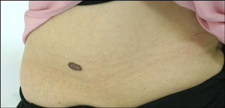
Fig. 1 Pruritic, solitary, well-demarcated, brown to black plaque, 3.8×1.2 cm in size, on the abdomen.
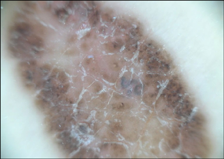
Fig. 2 Dermoscopic finding of the lesion showed a globular pattern with brown dots and globules at the periphery.
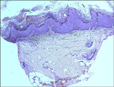
Fig. 3 Hyperkeratosis, parakeratosis, acanthosis, papillomatosis and hypergranulosis in the epidermis which was consistent with seborrheic keratosis (H&E, ×40).
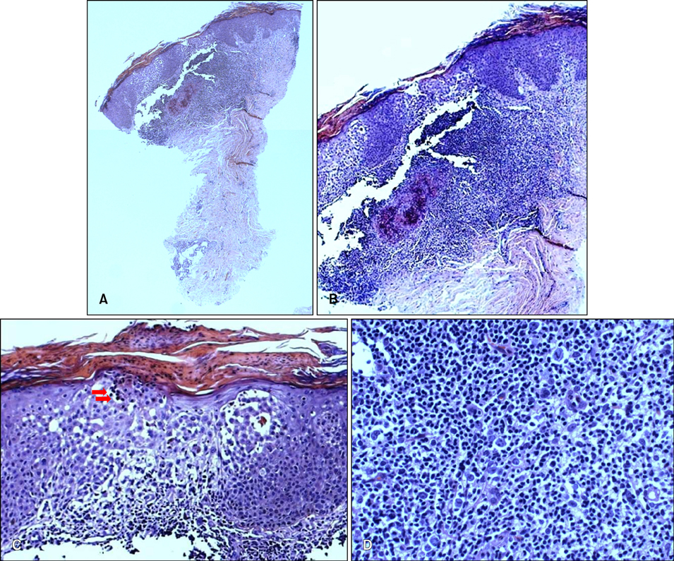
Fig. 4 (A, B) Epidermal hyperplasia and epidermotropism in the epidermis and formation of lymphoid follicle and lichenoid infiltration of lymphocytes in the reticular dermis (H&E stain, ×40, ×100). (C) Epidermotropism with Pautrier's microabscess in the epidermis (H&E stain, ×400). (D) Lichenoid infiltration of lymphocytes in the reticular dermis (H&E stain, ×400).
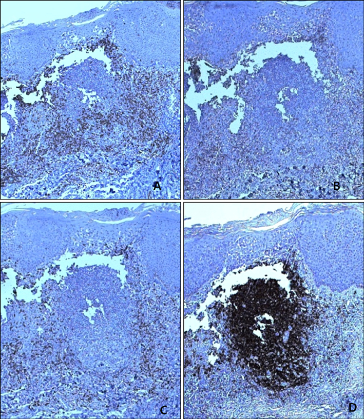
Fig. 5 Immunohistochemical study for CD3 (A), CD4 (B), CD8 (C), CD20 (D), showed positive staining with lymphocytes (H&E, ×400).
Reference
-
1. Evans LT, Mackey SL, Vidmar DA. An asymptomatic scaly plaque. Unilesional mycosis fungoides (MF). Arch Dermatol. 1997. 133:231. 234.2. Kossard S. Unilesional mycosis fungoides or lymphomatoid keratosis? Arch Dermatol. 1997. 133:1312–1313.
Article3. Arai E, Shimizu M, Tsuchida T, Izaki S, Ogawa F, Hirose T. Lymphomatoid keratosis: an epidermotropic type of cutaneous lymphoid hyperplasia: clinicopathological, immunohistochemical, and molecular biological study of 6 cases. Arch Dermatol. 2007. 143:53–59.4. Al-Hoqail IA, Crawford RI. Benign lichenoid keratoses with histologic features of mycosis fungoides: clinicopathologic description of a clinically significant histologic pattern. J Cutan Pathol. 2002. 29:291–294.
Article5. Lever WF, Schaumburg-Lever G. Histopathology of the skin. 1983. 6th ed. London: Lippincott;762–768.6. Bergman R, Khamaysi Z, Sahar D, Ben-Arieh Y. Cutaneous lymphoid hyperplasia presenting as a solitary facial nodule: clinical, histopathological, immunophenotypical, and molecular studies. Arch Dermatol. 2006. 142:1561–1566.7. Heald PW, Glusac EJ. Unilesional cutaneous T-cell lymphoma: clinical features, therapy, and follow-up of 10 patients with a treatment-responsive mycosis fungoides variant. J Am Acad Dermatol. 2000. 42:283–285.
Article8. Murphy GF, Schwarting R. Elder DE, Elenitsas R, Johnson BL, Murphy GF, editors. Cutaneous lymphomas and leukemias. Lever's histopathology of the skin. 2005. 9th ed. Philadelphia: Lippincott Williams & Wilkins;927–978.9. Bazza MA, Ryatt KS, Dharmagunawardena PV. Mycosis fungoides masquerading as seborrhoeic keratosis. Br J Dermatol. 2002. 147:1264–1265.
Article10. Yoo SS, Viglione M, Moresi M, Vonderheid E. Unilesional mycosis fungoides mimicking Bowen's disease. J Dermatol. 2003. 30:417–419.
Article
