Ann Dermatol.
2012 Aug;24(3):376-379. 10.5021/ad.2012.24.3.376.
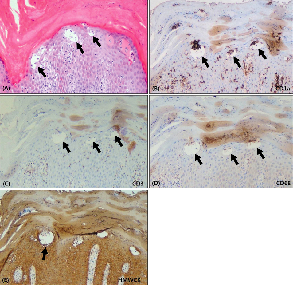
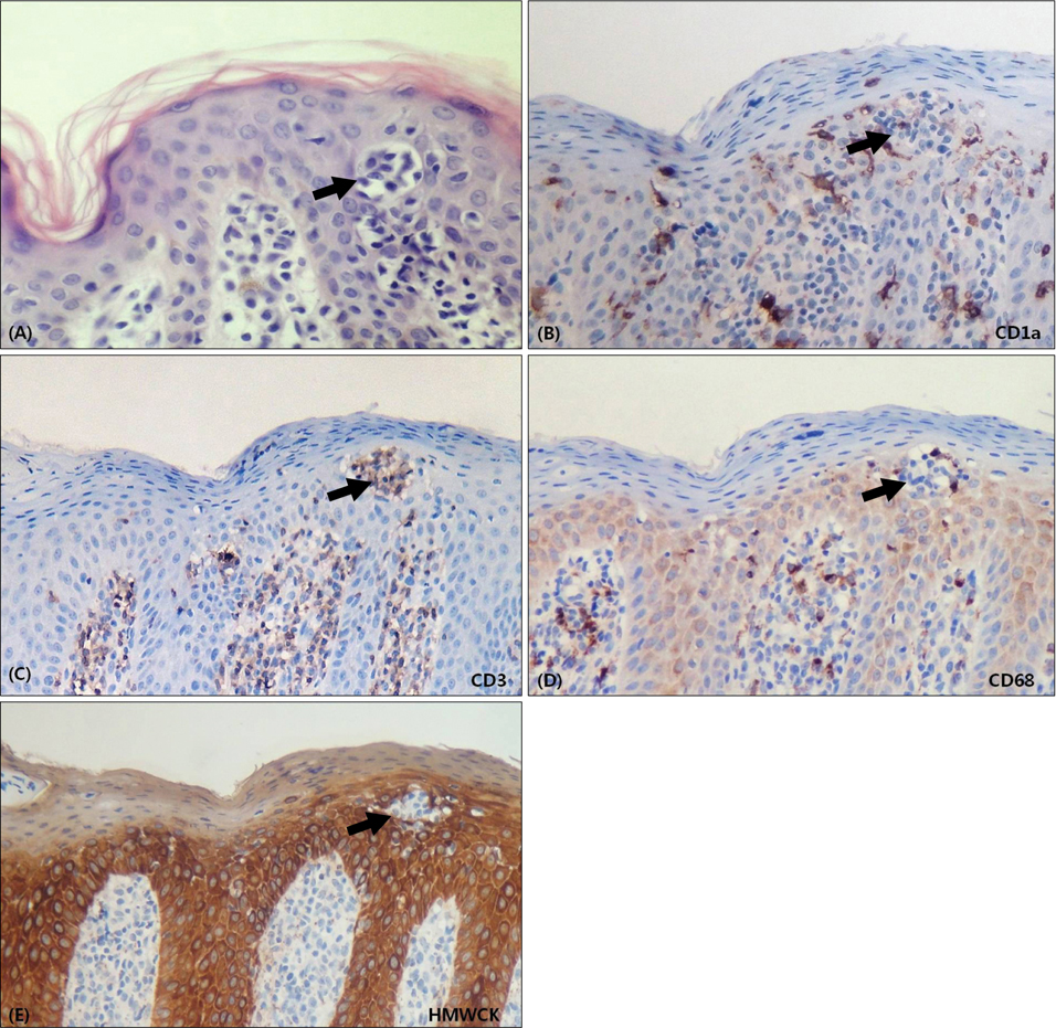
Comment on "Pseudopautrier's Abscess"
- Affiliations
-
- 1Department of Dermatology, Kosin University College of Medicine, Busan, Korea. ksderm98@unitel.co.kr
- KMID: 2265320
- DOI: http://doi.org/10.5021/ad.2012.24.3.376
Abstract
- No abstract available.
Figure
Reference
-
1. Lee SY, Kwon HC, Cho YS, Nam KH, Ihm CW, Kim JS. The three dimensional conformal radiotherapy for hyperkeratotic plantar mycosis fungoides. Ann Dermatol. 2011. 23:Suppl 1. S57–S60.
Article2. Girardi M, Heald PW, Wilson LD. The pathogenesis of mycosis fungoides. N Engl J Med. 2004. 350:1978–1988.
Article3. Ackerman AB, Breza TS, Capland L. Spongiotic simulants of mycosis fungoides. Arch Dermatol. 1974. 109:218–220.
Article4. Candiago E, Marocolo D, Manganoni MA, Leali C, Facchetti F. Nonlymphoid intraepidermal mononuclear cell collections (pseudo-Pautrier abscesses): a morphologic and immunophenotypical characterization. Am J Dermatopathol. 2000. 22:1–6.5. Kang DY, Baek JW, Jang MS, Suh KS, Kim ST. Expression patterns of pseudopautrier abscesses in mycosis fungoides. Korean J Dermatol. 2011. 49:Suppl 2. 180–181.
Article
- Full Text Links
- Actions
-
Cited
- CITED
-
- Close
- Share
- Similar articles
-
- Reply to the Comment on Adoption of Artificial Intelligence, Preprints, Open Peer Review, Model Text Recycling Policies, Best Practice in Scholarly Publishing: Comment
- Clinical Observation of Brodie's Abscess
- A Case of Frontal Sinus Osteoma Causing Brain Abscess
- A Case of Klebsiella Pneuomoniae Liver Abscess Complicated with Brain Abscess and Endophthalmitis
- Case Report of Treatment of IVlultiloculated Liver Abscess: Administration of Urokinase Through Drainage Catheter


