Ann Dermatol.
2015 Feb;27(1):92-94. 10.5021/ad.2015.27.1.92.
Adult Onset Dyschomatosis Universalis
- Affiliations
-
- 1Department of Dermatology, School of Medicine, Chosun University, Gwangju, Korea. derm75@chosun.ac.kr
- KMID: 2264846
- DOI: http://doi.org/10.5021/ad.2015.27.1.92
Abstract
- No abstract available.
Figure
-
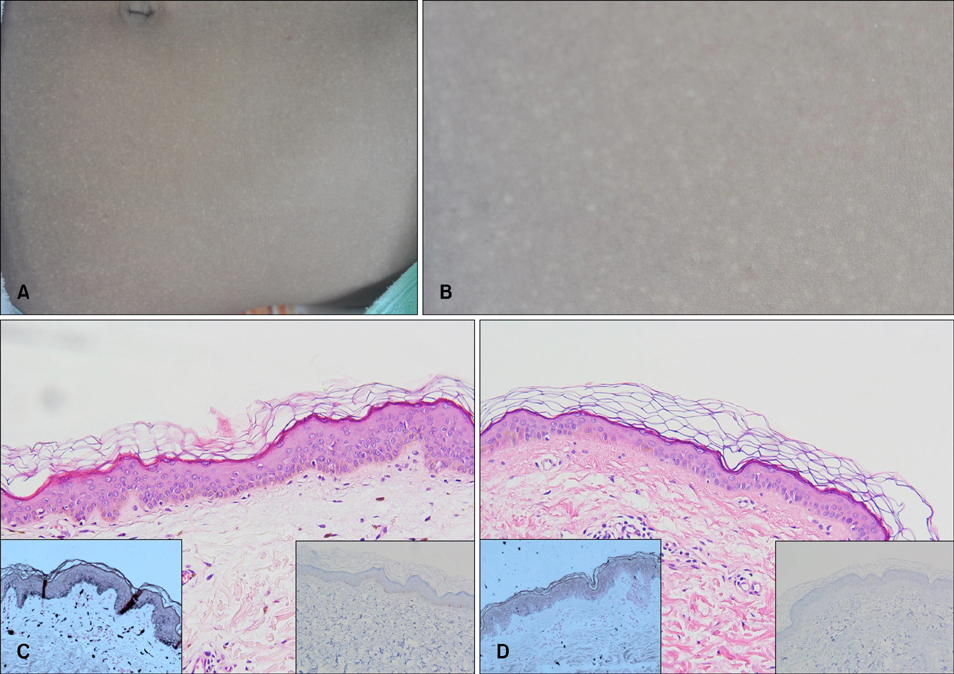
Fig. 1 (A) Multiple pinhead to rice sized mottled hypopigmented macules with diffuse hyperpigmented patches. (B) A closer view. (C) A biopsy specimen taken from a hyperpigmented lesion on the abdomen showed increased abundant melanin pigments in the epidermis with a normal number and distribution of melanocytes (H&E, ×200; left inset: Fontana Masson, ×200; right inset: melanoma antigen recognized by T cells 1 [MART-1] ×200). (D) A hypopigmented macule showed reduced amount of melanin pigments in the lesion area (H&E, ×200; left inset: Fontana Masson, ×200; right inset: MART-1, ×200).
Reference
-
1. Urabe K, Hori Y. Dyschromatosis. Semin Cutan Med Surg. 1997; 16:81–85.
Article2. Kim YJ, Oh CN, Chung BS, Choi KC. Two cases of dyschromatosis universalis. Korean J Dermatol. 1992; 30:928–931.3. Rycroft RJ, Calnan CD, Wells RS. Universal dyschromatosis, small stature and high-tone deafness. Clin Exp Dermatol. 1977; 2:45–48.
Article4. Shono S, Toda K. Universal dyschromatosis associated with photosensitivity and neurosensory hearing defect. Arch Dermatol. 1990; 126:1659–1660.
Article5. Kim NS, Im S, Kim SC. Dyschromatosis universalis hereditaria: an electron microscopic examination. J Dermatol. 1997; 24:161–164.
Article
- Full Text Links
-
- Actions
-
Cited
- CITED
-
- Close
- Share
-
- Similar articles
-
- A Case of Alopecia Universalis in a Gravida
- Clinical Characteristics and Prognostic Factors in Early-Onset Alopecia Totalis and Alopecia Universalis
- Dyschromatosis Universalis Hereditaria
- Systematized Epidermal Nevi Associated with Congenital Alopecia Universalis and Onychodystrophy
- Vitiligo Universalis Associated with Chronic Hepatitis B
