J Adv Prosthodont.
2014 Apr;6(2):71-78. 10.4047/jap.2014.6.2.71.
Evaluation of the color reproducibility of all-ceramic restorations fabricated by the digital veneering method
- Affiliations
-
- 1Department of Dental Laboratory Science and Engineering, College of Health Science & Department of Public Health Sciences, Graduate School, Korea University, Seoul, Republic of Korea. kjh2804@korea.ac.kr
- 2Department of Dental Laboratory Science and Engineering, College of Health Science & Department of Public Health Sciences, Graduate School & BK21+ Program in Public Health Sciences, Korea University, Seoul, Republic of Korea.
- KMID: 2233333
- DOI: http://doi.org/10.4047/jap.2014.6.2.71
Abstract
- PURPOSE
The objective of this study was to evaluate the clinical acceptability of all-ceramic crowns fabricated by the digital veneering method vis-a-vis the traditional method.
MATERIALS AND METHODS
Zirconia specimens manufactures by two different manufacturing method, conventional vs digital veneering, with three different thickness (0.3 mm, 0.5 mm, 0.7 mm) were prepared for analysis. Color measurement was performed using a spectrophotometer for the prepared specimens. The differences in shade in relation to the build-up method were calculated by quantifying DeltaE* (mean color difference), with the use of color difference equations representing the distance from the measured values L*, a*, and b*, to the three-dimensional space of two colors. Two-way analysis of variance (ANOVA) combined with a Tukey multiple-range test was used to analyze the data (alpha=0.05).
RESULTS
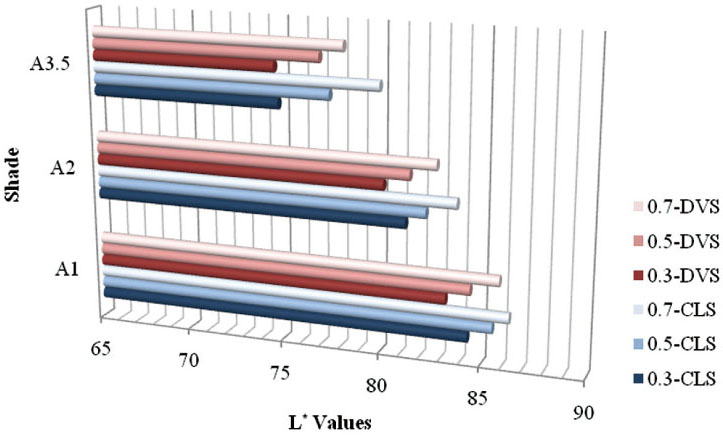
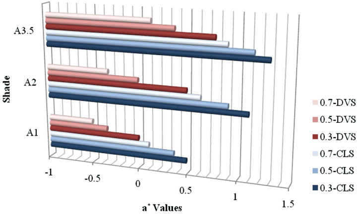
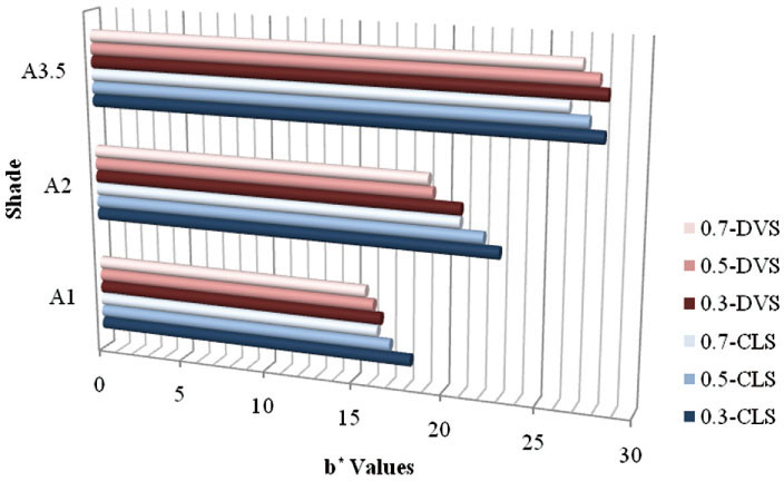
In comparing means and standard deviations of L*, a*, and b* color values there was no significant difference by the manufacturing method and zirconia core thickness according to a two-way ANOVA. The color differences between two manufacturing methods were in a clinically acceptable range less than or equal to 3.7 in all the specimens.
CONCLUSION
Based on the results of this study, a carefully consideration is necessary while selecting upper porcelain materials, even if it is performed on a small scale. However, because the color reproducibility of the digital veneering system was within the clinically acceptable range when comparing with conventional layering system, it was possible to estimate the possibility of successful aesthetic prostheses in the latest technology.
Figure
Reference
-
1. Preston JD. Current status of shade selection and color matching. Quintessence Int. 1985; 16:47–58.2. Dozić A, Kleverlaan CJ, Meegdes M, van der Zel J, Feilzer AJ. The influence of porcelain layer thickness on the final shade of ceramic restorations. J Prosthet Dent. 2003; 90:563–570.3. Seghi RR, Johnston WM, O'Brien WJ. Spectrophotometric analysis of color differences between porcelain systems. J Prosthet Dent. 1986; 56:35–40.4. O'Brien WJ, Kay KS, Boenke KM, Groh CL. Sources of color variation on firing porcelain. Dent Mater. 1991; 7:170–173.5. Guazzato M, Albakry M, Ringer SP, Swain MV. Strength, fracture toughness and microstructure of a selection of all-ceramic materials. Part II. Zirconia-based dental ceramics. Dent Mater. 2004; 20:449–456.6. Braga RR, Ballester RY, Daronch M. Influence of time and adhesive system on the extrusion shear strength between feldspathic porcelain and bovine dentin. Dent Mater. 2000; 16:303–310.7. Aboushelib MN, Kleverlaan CJ, Feilzer AJ. Effect of zirconia type on its bond strength with different veneer ceramics. J Prosthodont. 2008; 17:401–408.8. Choi YS, Kim SH, Lee JB, Han JS, Yeo IS. In vitro evaluation of fracture strength of zirconia restoration veneered with various ceramic materials. J Adv Prosthodont. 2012; 4:162–169.9. Hochman N, Zalkind M. New all-ceramic indirect post-and-core system. J Prosthet Dent. 1999; 81:625–629.10. Douglas RD, Przybylska M. Predicting porcelain thickness required for dental shade matches. J Prosthet Dent. 1999; 82:143–149.11. Heffernan MJ, Aquilino SA, Diaz-Arnold AM, Haselton DR, Stanford CM, Vargas MA. Relative translucency of six all-ceramic systems. Part I: core materials. J Prosthet Dent. 2002; 88:4–9.12. O'Brien WJ, Groh CL, Boenke KM. A new, small-color-difference equation for dental shades. J Dent Res. 1990; 69:1762–1764.13. Johnston WM, Kao EC. Assessment of appearance match by visual observation and clinical colorimetry. J Dent Res. 1989; 68:819–822.14. Shotwell JL, Razzoog ME, Koran A. Color stability of long-term soft denture liners. J Prosthet Dent. 1992; 68:836–838.15. Douglas RD, Steinhauer TJ, Wee AG. Intraoral determination of the tolerance of dentists for perceptibility and acceptability of shade mismatch. J Prosthet Dent. 2007; 97:200–208.16. Preston JD. Current status of shade selection and color matching. Quintessence Int. 1985; 16:47–58.17. Kelly JR, Nishimura I, Campbell SD. Ceramics in dentistry: historical roots and current perspectives. J Prosthet Dent. 1996; 75:18–32.18. Knispel G. Factors affecting the process of color matching restorative materials to natural teeth. Quintessence Int. 1991; 22:525–531.19. Fasbinder DJ. Digital dentistry: innovation for restorative treatment. Compend Contin Educ Dent. 2010; 31:2–11. quiz 12.20. Luo XP, Zhang L. Effect of veneering techniques on color and translucency of Y-TZP. J Prosthodont. 2010; 19:465–470.21. Lee YK, Cha HS, Ahn JS. Layered color of all-ceramic core and veneer ceramics. J Prosthet Dent. 2007; 97:279–286.22. Lund PS, Piotrowski TJ. Color changes of porcelain surface colorants resulting from firing. Int J Prosthodont. 1992; 5:22–27.
- Full Text Links
- Actions
-
Cited
- CITED
-
- Close
- Share
- Similar articles
-
- Color stability of fully- and pre-crystalized chair-side CAD-CAM lithium disilicate restorations after required and additional sintering processes
- Color stability of fully- and pre-crystalized chair-side CAD-CAM lithium disilicate restorations after required and additional sintering processes
- Effect of the shades of background substructures on the overall color of zirconia-based all-ceramic crowns
- Full-mouth rehabilitation with pressed ceramic technique using provisional restorations
- Spectrophotometric analysis of the influence of zirconia core on the color of ceramic


