Imaging Sci Dent.
2013 Dec;43(4):283-287. 10.5624/isd.2013.43.4.283.
Prevalence and characteristics of pneumatized articular tubercle: First large series in Iranian people
- Affiliations
-
- 1Department of Oral and Maxillofacial Radiology, Dental School, Hamadan University of Medical Sciences, Hamadan, Iran.
- 2Department of Oral and Maxillofacial Medicine, Dental School, Shahid Beheshti University of Medical Sciences, Tehran, Iran. hamedmortazavi2013@gmail.com
- KMID: 2229614
- DOI: http://doi.org/10.5624/isd.2013.43.4.283
Abstract
- PURPOSE
This study was performed to determine the prevalence and characteristics of pneumatized articular tubercle or eminence among a defined group of Iranian people.
MATERIALS AND METHODS
Digital panoramic radiographs of 1694 patients in the Department of Oral and Maxillofacial Radiology, Hamadan Dental School, Iran were evaluated retrospectively to detect the above lesion. Finally, 1563 radiographs were selected according to inclusion criteria. Then, a review was done of 10 large case series found using a MEDLINE search of the literature. Chi-squared test was used to analyze the differences in variables such as age, gender, laterality, and locularity in our case series.
RESULTS
The average age of our samples was 32.6+/-7.63 years. Pneumatized articular tubercle was found in 98 cases, representing a prevalence of 6.2% with a mean age of 22.8+/-7.9 and a range of 8 to 60 years. Sixty-four (65.3%) pneumatized articular tubercles were unilateral, with 30 lesions on the right and 34 on the left side. Bilateral lesions were found in 34 (34.7%) patients. 52 (53.06%) of the pneumatized articular tubercles were of the unilocular type and 46 (46.94%) were multilocular. The results showed no statistically significant differences regarding age (p=0.454), gender (p=0.634), laterality (p=0.252), or locularity (p=0.807) among the samples.
CONCLUSION
Among ten large case series from other countries, the prevalence of pneumatized articular tubercle (6.2%) in Iranian patients was higher than that of all eight of the case series that used the same detection method as the present study of panoramic radiography.
Keyword
MeSH Terms
Figure
-
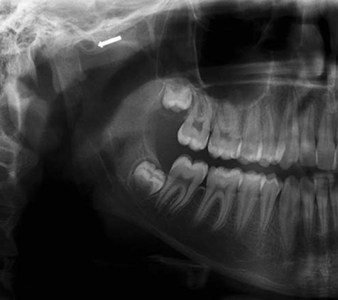
Fig. 1 A cropped panoramic radiograph shows a unilocular pneumatized articular tubercle.
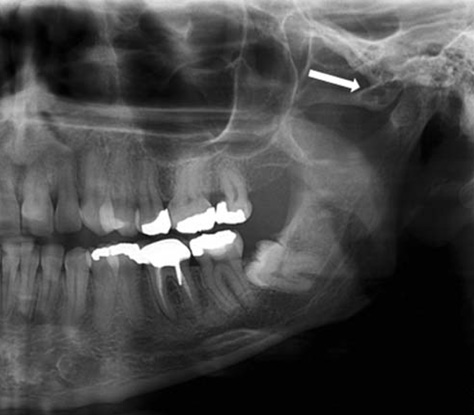
Fig. 2 A cropped panoramic radiograph shows a multilocular pneumatized articular tubercle.
Reference
-
1. Stoopler ET, Pinto A, Stanton DC, Mupparapu M, Sollecito TP. Extensive pneumatization of the temporal bone and articular eminence: an incidental finding in a patient with facial pain. Case report and review of literature. Quintessence Int. 2003; 34:211–214.2. Deluke DM. Pneumatization of the articular eminence of the temporal bone. Oral Surg Oral Med Oral Pathol Oral Radiol Endod. 1995; 79:3–4.
Article3. Sözen E, Çelebi I, Uçal YO, Coşkun BU. Is there a relationship between subjective pulsatile tinnitus and petrous bone pneumatization? J Craniofac Surg. 2013; 24:461–463.4. Tyndall DA, Matteson SR. Radiographic appearance and population distribution of the pneumatized articular eminence of the temporal bone. J Oral Maxillofac Surg. 1985; 43:493–497.
Article5. Carter LC, Haller AD, Calamel AD, Pfaffenbach AC. Zygomatic air cell defect (ZACD). Prevalence and characteristics in a dental clinic outpatient population. Dentomaxillofac Radiol. 1999; 28:116–122.
Article6. Ladeira DB, Barbosa GL, Nascimento MC, Cruz AD, Freitas DQ, Almeida SM. Prevalence and characteristics of pneumatization of the temporal bone evaluated by cone beam computed tomography. Int J Oral Maxillofac Surg. 2013; 42:771–775.
Article7. Hasnaini M, Ng SY. Extensive temporal bone pneumatization: incidental finding in a patient with TMJ dysfunction. Dent Update. 2000; 27:187–189.
Article8. Miloglu O, Yilmaz AB, Yildirim E, Akgul HM. Pneumatization of the articular eminence on cone beam computed tomography: prevalence, characteristics and a review of the literature. Dentomaxillofac Radiol. 2011; 40:110–114.
Article9. Kaugars GE, Mercuri LG, Laskin DM. Pneumatization of the articular eminence of the temporal bone: prevalence, development, and surgical treatment. J Am Dent Assoc. 1986; 113:55–57.
Article10. Hofmann T, Friedrich RE, Wedl JS, Schmelzle R. Pneumatization of the zygomatic arch on pantomography. Mund Kiefer Gesichtschir. 2001; 5:173–179.11. Orhan K, Delilbasi C, Cebeci I, Paksoy C. Prevalence and variations of pneumatized articular eminence: a study from Turkey. Oral Surg Oral Med Oral Pathol Oral Radiol Endod. 2005; 99:349–354.
Article12. Orhan K, Delilbasi C, Orhan AI. Radiographic evaluation of pneumatized articular eminence in a group of Turkish children. Dentomaxillofac Radiol. 2006; 35:365–370.
Article13. Yavuz MS, Aras MH, Güngör H, Büyükkurt MC. Prevalence of the pneumatized articular eminence in the temporal bone. J Craniomaxillofac Surg. 2009; 37:137–139.
Article14. Orhan K, Oz U, Orhan AI, Ulker AE, Delilbasi C, Akcam O. Investigation of pneumatized articular eminence in orthodontic malocclusions. Orthod Craniofac Res. 2010; 13:56–60.
Article15. Roser SM, Rudin DE, Brady FA. Unusual bony lesion of the zygomatic arch. J Oral Med. 1976; 31:72–73.
- Full Text Links
-
- Actions
-
Cited
- CITED
-
- Close
- Share
-
- Similar articles
-
- Comparison of mastoid air cell volume in patients with or without a pneumatized articular tubercle
- Knowledge of and attitudes toward HIV/AIDS among Iranian women
- A vertically split fracture of the marginal tubercle of the zygoma in a 3-year-old boy: a case report
- Epidemiology of Rheumatoid Arthritis in Korea
- A Clinical Study of Tubercle Bacilli in Urine of the Patients with Pulmonary Tuberculosis
