Posterior Reversible Encephalopathy Syndrome in a Critically Ill Postoperative Patient
- Affiliations
-
- 1Division of Trauma and Surgical Critical Care, Department of Surgery, University of Ulsan College of Medicine, Asan Medical Center, Seoul, Korea. skhong94@amc.seoul.kr
- 2Department of Radiology and Research Institute of Radiology, Asan Medical Center, University of Ulsan College of Medicine, Seoul, Korea.
- 3Department of Neurology, Asan Medical Center, University of Ulsan College of Medicine, Seoul, Korea.
- KMID: 2227702
- DOI: http://doi.org/10.4266/kjccm.2015.30.1.46
Abstract
- Posterior reversible encephalopathy syndrome (PRES) is a transient condition characterized by altered mental status, seizure, headache, and visual disturbance with typical neuro-imaging findings in the bilateral parieto-occipital regions. Clinicians should be aware of this syndrome because delayed diagnosis and treatment result in irreversible neurologic deficits. We present the case of a 77-year-old male diagnosed with PRES in the setting of postoperative critical illness caused by small-bowel strangulation.
MeSH Terms
Figure
-
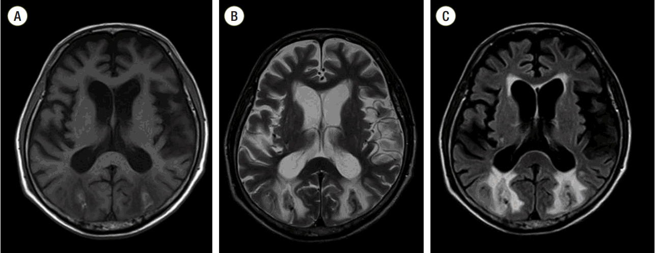
Fig. 1. MRI assessment of chronic changes related to PRES with internal intracerebral hemorrhage. Axial T1-weighted imaging (A) and axial T2-weighted imaging (B) revealed subacute intracerebral hemorrhages in the bilateral occipital lobes, which were seen as high signal intensities on T1-weighted imaging and as a dark rim on T2-weighted imaging. In the deep and subcortical white matter of the bilateral occipital lobes, cerebral edema was also seen as high signal intensities on T2-weighted (B) and fluid-attenuated inversion recovery imaging (C). MRI: magnetic resonance imaging; PRES: posterior reversible encephalopathy syndrome.
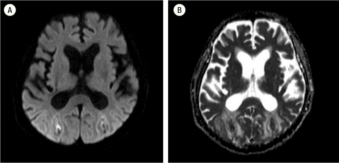
Fig. 2. MRI assessment of chronic change related to PRES with internal intracerebral hemorrhage. DWI (A) and ADC map imaging (B) did not reveal significant signal changes in the corresponding areas, except in the hemorrhagic foci. MRI: magnetic resonance imaging; PRES: posterior reversible encephalopathy syndrome; DWI: diffusion weighted imaging; ADC: apparent diffusion coefficient.
Reference
-
References
1. Hinchey J, Chaves C, Appignani B, Breen J, Pao L, Wang A, et al. A reversible posterior leukoencephalopathy syndrome. N Engl J Med. 1996; 334:494–500.
Article2. Bartynski WS. Posterior reversible encephalopathy syndrome, part 1: fundamental imaging and clinical features. AJNR Am J Neuroradiol. 2008; 29:1036–42.
Article3. Bartynski WS. Posterior reversible encephalopathy syndrome, part 2: controversies surrounding pathophysiology of vasogenic edema. AJNR Am J Neuroradiol. 2008; 29:1043–9.
Article4. Bartynski WS, Boardman JF. Distinct imaging patterns and lesion distribution in posterior reversible encephalopathy syndrome. AJNR Am J Neuroradiol. 2007; 28:1320–7.
Article5. Roth C, Ferbert A. The posterior reversible encephalopathy syndrome: what’s certain, what’s new? Pract Neurol. 2011; 11:136–44.
Article6. Casey SO, Sampaio RC, Michel E, Truwit CL. Posterior reversible encephalopathy syndrome: utility of fluid-attenuated inversion recovery MR imaging in the detection of cortical and subcortical lesions. AJNR Am J Neuroradiol. 2000; 21:1199–206.7. Lee VH, Wijdicks EF, Manno EM, Rabinstein AA. Clinical spectrum of reversible posterior leukoencephalopathy syndrome. Arch Neurol. 2008; 65:205–10.
Article8. Fugate JE, Claassen DO, Cloft HJ, Kallmes DF, Kozak OS, Rabinstein AA. Posterior reversible encephalopathy syndrome: associated clinical and radiologic findings. Mayo Clin Proc. 2010; 85:427–32.
Article9. Roth C, Ferbert A. Posterior reversible encephalopathy syndrome: long-term follow-up. J Neurol Neurosurg Psychiatry. 2010; 81:773–7.
Article10. Stott VL, Hurrell MA, Anderson TJ. Reversible posterior leukoencephalopathy syndrome: a misnomer reviewed. Intern Med J. 2005; 35:83–90.
Article11. Kwon S, Koo J, Lee S. Clinical spectrum of reversible posterior leukoencephalopathy syndrome. Pediatr Neurol. 2001; 24:361–4.
Article12. Ahn KJ, You WJ, Jeong SL, Lee JW, Kim BS, Lee JH, et al. Atypical manifestations of reversible posterior leukoencephalopathy syndrome: findings on diffusion imaging and ADC mapping. Neuroradiology. 2004; 46:978–83.
Article13. Servillo G, Bifulco F, De Robertis E, Piazza O, Striano P, Tortora F, et al. Posterior reversible encephalopathy syndrome in intensive care medicine. Intensive Care Med. 2007; 33:230–6.
Article14. Vaughan CJ, Delanty N. Hypertensive emergencies. Lancet. 2000; 356:411–7.
Article
- Full Text Links
-
- Actions
-
Cited
- CITED
-
- Close
- Share
-
- Similar articles
-
- Posterior Reversible Encephalopathy Syndrome in a Patient with Intoxication of Arisaema amurense
- Posterior Reversible Encephalopathy Syndrome after Massive Blood Transfusion in a Normotensive Patient
- Posterior reversible encephalopathy syndrome and reversible cerebral vasoconstriction syndrome associated with acute exacerbation of chronic obstructive pulmonary disease
- A Case of Posterior Reversible Encephalopathy Syndrome in a Patient having Continuous Ambulatory Peritoneal Dialysis
- Posterior Reversible Encephalopathy after Quetiapine Overdose
