Persistent Left Superior Vena Cava Detected Incidentally after Pulmonary Artery Catheterization
- Affiliations
-
- 1Department of Anesthesiology and Pain Medicine, Yonsei University College of Medicine, Seoul, Korea. SJW72331@yuhs.ac
- 2Anesthesia and Pain Research Institute, Yonsei University College of Medicine, Seoul, Korea.
- KMID: 2227696
- DOI: http://doi.org/10.4266/kjccm.2015.30.1.22
Abstract
- We present a case of pulmonary artery catheter (PAC) placement through the right internal jugular vein, bridging vein and coronary sinus in a patient with previously unrecognized persistent left superior vena cava (LSVC) and diminutive right superior vena cava. A 61-year-old male patient was scheduled for mitral valve repair for regurgitation. Preoperative transthoracic echocardiography revealed dilated coronary sinus, but no further evaluations were performed. During advancement of the PAC, right ventricular and pulmonary arterial pressure tracing was observed at 50 and 60 cm, respectively. Transesophageal echocardiography ruled out intracardiac knotting and revealed the presence of the PAC in the LSVC, entering the right ventricle from the coronary sinus. Diminutive right superior vena cava was observed after sternotomy. The PAC was left in place for 2 days postoperatively without any complications. This case emphasizes that the possibility of LSVC and associated anomalies should always be ruled out in patients with dilated coronary sinus.
MeSH Terms
Figure
-

Fig. 1. Transesophageal echocardiography revealed the presence of the pulmonary artery catheter (white arrow) in the left superior vena cava (A) and coronary sinus (B). LA: left atrium; LV: left ventricle; LSVC: left superior vena cava; TEE: transthoracic echocardiography; CS: coronary sinus.
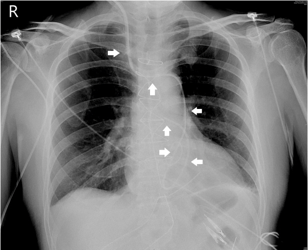
Fig. 2. Postoperative chest x-ray showed the unusual path of the pulmonary artery catheter (white arrows), suggesting that the catheter passed through the right internal jugular vein, bridging vein, left superior vena cava, coronary sinus, right ventricle and pulmonary artery. R: right.
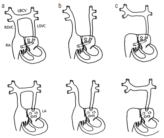
Fig. 3. Various presentations of persistent left superior vena cava. LBCV: left brachiocephalic vein; RSVC: right superior vena cava; LSVC: left superior vena cava; RA: right atrium; LA: left atrium.
Reference
-
References
1. Giuliani-Poncini C, Perez MH, Cotting J, Hurni M, Sekarski N, Pfammatter JP, et al. Persistent left superior vena cava in cardiac congenital surgery. Pediatr Cardiol. 2014; 35:71–6.
Article2. Erdoğan M, Karakaş P, Uygur F, Meşe B, Yamak B, Bozkir MG. Persistent left superior vena cava: the anatomical and surgical importance. West Indian Med J. 2007; 56:72–6.
Article3. Voci P, Luzi G, Agati L. Diagnosis of persistent left superior vena cava by multiplane transesophageal echocardiography. Cardiologia. 1995; 40:273–5.4. Goyal SK, Punnam SR, Verma G, Ruberg FL. Persistent left superior vena cava: a case report and review of literature. Cardiovasc Ultrasound. 2008; 6:50.
Article5. Konvicka JJ, Villamaria FJ. Images in anesthesia: anesthetic implications of persistent left superior vena cava. Can J Anaesth. 2005; 52:805.
Article6. Nsah EN, Moore GW, Hutchins GM. Pathogenesis of persistent left superior vena cava with a coronary sinus connection. Pediatr Pathol. 1991; 11:261–9.
Article7. Oosawa M, Sakai A, Abe M, Hanayama N, Lin ZB, Kodera K. Repeat open heart surgery in a case associated with persistent left superior vena cava: a method of simple occlusion of L-SVC using an alternative extra-pericardial approach and retrograde cardioplegia. Kyobu Geka. 1995; 48:741–4.8. Dhar P, Kaufman B, Doerfler M, Dadic P. Unusual course of a pulmonary artery catheter. J Cardiothorac Vasc Anesth. 1998; 12:487–9.
Article9. Huang SK. Persistent left superior vena cava in a man with ventricular fibrillation. Chest. 1986; 89:155–7.
Article10. Menéndez B, García del Valle S, Marcos RC, Azofra J, Gomez-Arnau J. Left superior vena cava: a vascular abnormality discovered following pulmonary artery catheterization. Can J Anaesth. 1996; 43:626–8.
Article11. Rose ME, Gross L, Protos A. Transvenous pacemaker implantation by way of an anomalous left superior vena cava. J Thorac Cardiovasc Surg. 1971; 62:965–6.
Article12. Roberts DH, Bellamy CM, Ramsdale DR. Implantation of a dual chamber pacemaker in a patient with persistent left superior vena cava. Int J Cardiol. 1992; 36:242–3.
Article13. Meijboom WB, Vanderheyden M. Biventricular pacing and persistent left superior vena cava. Case report and review of the literature. Acta Cardiol. 2002; 57:287–90.
- Full Text Links
-
- Actions
-
Cited
- CITED
-
- Close
- Share
-
- Similar articles
-
- A Case of Persistent Left Superior Vena Cava with Interruption of Inferior Vena Cava
- Persistent Left Superior Vena Cava with Absent Right Superior Vena Cava and Large Atrial Septal Defect in Visceroatrial Situs solitus
- Persistent Left Sperior Vena Cava Draining into the Left Atrium with Absent Right Superior Vena Cava in Tetralogy of Fallot
- Congenital Absence of the Azygos Vein with Persistent Left Superior Vena Cava: A Case Report
- Pulmonary artery sling: case report
