Korean Circ J.
2007 Dec;37(12):666-670. 10.4070/kcj.2007.37.12.666.
Transthoracic Echocardiographic Detection, Differential Diagnosis, and Follow-Up of Esophageal Hematoma
- Affiliations
-
- 1Division of Cardiology, Cardiovascular Center, Yonsei University College of Medicine, Seoul, Korea. namsikc@yuhs.ac
- KMID: 2227044
- DOI: http://doi.org/10.4070/kcj.2007.37.12.666
Abstract
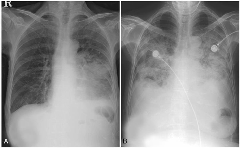
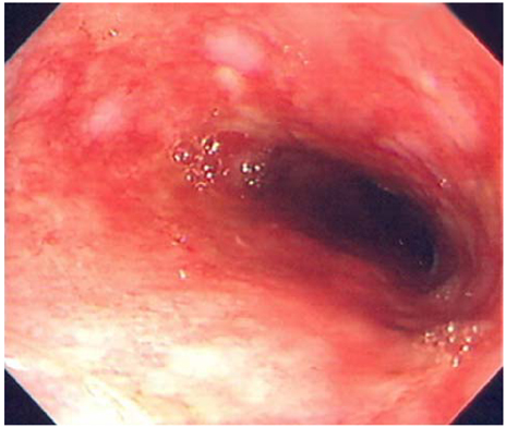
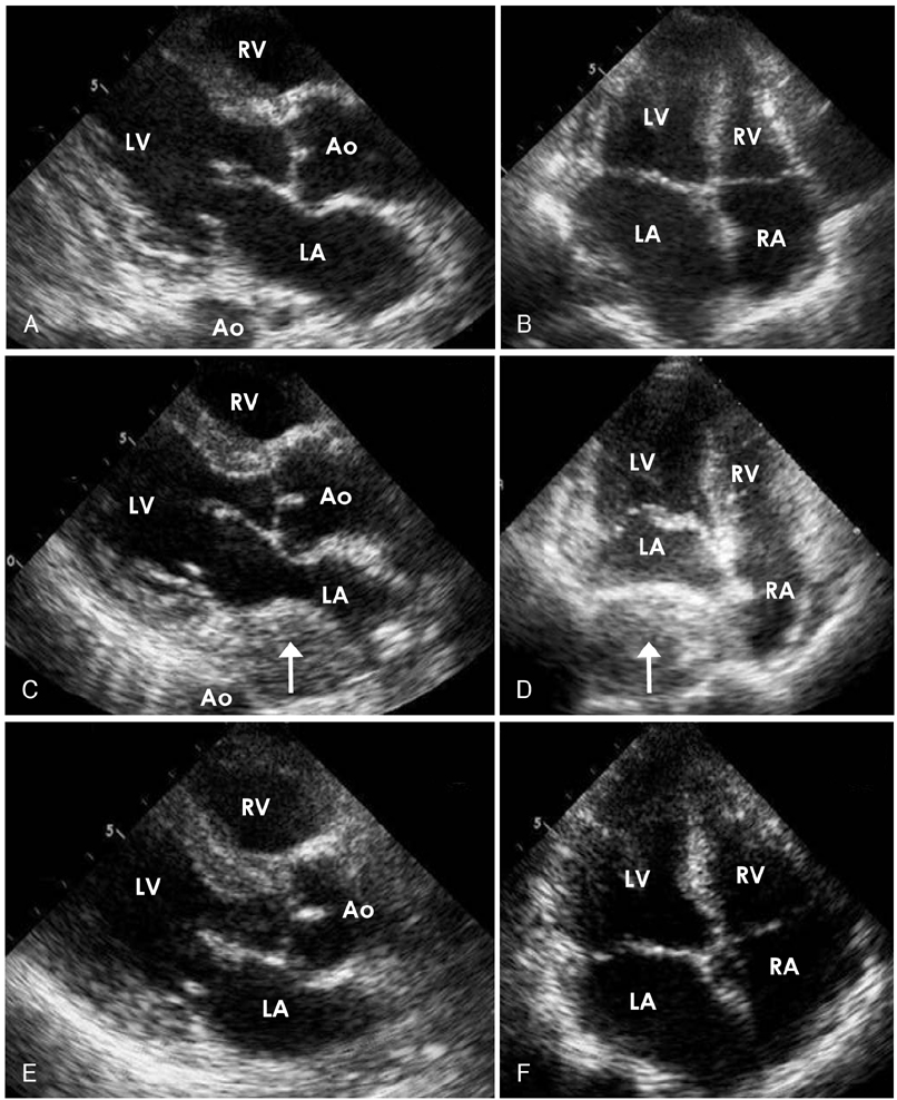
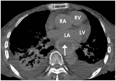
- Esophageal hematoma is a rare form of esophageal injury. It may occur spontaneously, or in association with direct esophageal damage or a bleeding diathesis. Endoscopy and computed tomography are generally necessary for the establishment of a diagnosis. In this report, we present a case of esophageal hematoma that was discovered via a bedside transthoracic echocardiography. The echocardiography was conducted to evaluate an unexplained shock in a critically ill-patient. After conservative treatment, complete resolution of the esophageal hematoma was documented by a 7-day short-term follow-up of bedside transthoracic echocardiography. To the best of our knowledge, this is the first case report regarding transthoracic echocardiographic detection, differential diagnosis, and follow-up for esophageal hematoma.
Keyword
MeSH Terms
Figure
Reference
-
1. Yamashita K, Okuda H, Fukushima H, Arimura Y, Endo T, Imai K. A case of intramural esophageal hematoma: complication of anticoagulation with heparin. Gastrointest Endosc. 2000. 52:559–561.2. Mosimann F, Bronnimann B. Intramural haematoma of the oesophagus complicating sclerotherapy for varices. Gut. 1994. 35:130–131.3. Ashman FC, Hill MC, Saba GP, Diaconis JN. Esophageal hematoma associated with thrombocytopenia. Gastrointest Radiol. 1978. 3:115–118.4. Enns R, Brown JA, Halparin L. Intramural esophageal hematoma: a diagnostic dilemma. Gastrointest Endosc. 2000. 51:757–759.5. Kim TW, Chang KS, Her GM, et al. Usefulness of echocardiography in the evaluation of paracardiac masses. Korean Circ J. 1996. 26:803–812.6. Williams B. Case report: oesophageal laceration following remote trauma. Br J Radiol. 1957. 30:666–668.7. Cullen SN, McIntyre AS. Dissecting intramural haematoma of the oesophagus. Eur J Gastroenterol Hepatol. 2000. 12:1151–1162.8. Phan GQ, Heitmiller RF. Intramural esophageal dissection. Ann Thorac Surg. 1997. 63:1785–1786.9. Reiter C, Denk W. Dissecting esophageal hematoma following thoracic aorta rupture. Unfallchirurg. 1985. 88:322–326.10. Hunter TB, Protell RL, Horsley WW. Food laceration of the esophagus: the taco tear. AJR Am J Roentgenol. 1983. 140:503–504.11. Criblez D, Filippini L, Schoch O, Meier UR, Koelz HR. Intramural rupture and intramural hematoma of the esophagus: 3 case reports and literature review. Schweiz Med Wochenschr. 1992. 122:416–423.12. Lu MS, Liu YH, Liu HP, Wu YC, Chu Y, Chu JJ. Spontaneous intramural esophageal hematoma. Ann Thorac Surg. 2004. 78:343–345.13. Cullen SN, Chapman RW. Dissecting intramural haematoma of the oesophagus exacerbated by heparin therapy. QJM. 1999. 92:123–124.14. Geller A, Gostout CJ. Esophagogastric hematoma mimicking a malignant neoplasm: clinical manifestations, diagnosis, and treatment. Mayo Clin Proc. 1998. 73:342–345.15. Herbetko J, Delany D, Ogilvie BC, Blaquiere RM. Spontaneous intramural haematoma of the oesophagus: appearance on computed tomography. Clin Radiol. 1991. 44:327–328.16. Kwak MH, Oh J, Jeong JO, et al. Role of echocardiography as a screening test in patients with suspected pulmonary embolism. Korean Circ J. 2001. 31:500–506.
- Full Text Links
- Actions
-
Cited
- CITED
-
- Close
- Share
- Similar articles
-
- Surgical Treatment of the Esophageal Diverticulum
- Echocardiographic Findings of Heart Disease in Children
- A Case of Obstructive Esophageal Hematoma after Endoscopic Variceal Ligation
- Unusual Diaphragmatic Hernias Mimicking Cardiac Masses
- Esophageal duplication cyst complicated with intramural hematoma: case report


