A Sirolimus: Eluting Stent Fracture Combined with a Coronary Artery Aneurysm
- Affiliations
-
- 1Division of Cardiology, Cardiovascular Center, Yonsei University College of Medicine, Seoul, Korea. whshim@yuhs.ac
- KMID: 2225877
- DOI: http://doi.org/10.4070/kcj.2008.38.1.69
Abstract
- A stent fracture combined with a coronary artery aneurysm is a rare event. As these events can lead to a harmful outcome, such as the development of myocardial ischemia by in-stent restenosis or thrombosis, repeated coronary intervention may be required. We report a case of a stent fracture combined with a coronary artery aneurysm. The fracture was thought to have developed by mechanical stress produced from a change of regional wall motion after an anteroseptal myocardial infarction. As detected by the use of intravascular ultrasound, neither in-stent restenosis nor a thrombus in the fractured stent was present. A cardiac magnetic resonance image showed that no viable myocardium in the anteroseptal wall was present. Therefore, the patient underwent medical treatment without intervention of the fractured stent.
Keyword
MeSH Terms
Figure
-
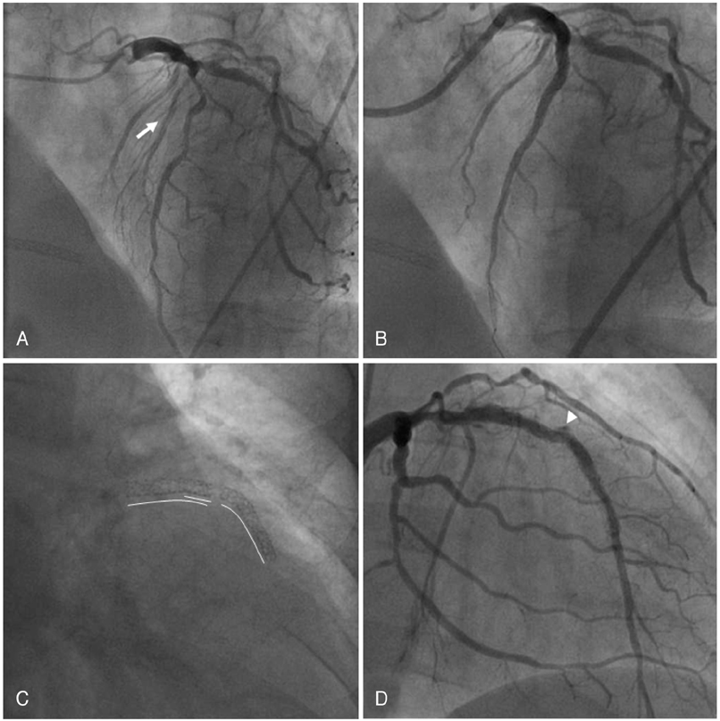
Fig. 1 Serial findings of coronary angiogram. A: initial coronary angiography showed diffuse stenosis in the proximal and mid portions of the LAD. B: post PCI angiography revealed well-deployed overlapping SESs, but the big septal artery (arrow) was totally occluded. C: fluoroscopy at follow-up showed a complete SES fracture. D: coronary angiography showed coronary artery aneurysm (arrowhead) at the fractured site. LAD: left anterior descending artery, PCI: percutaneous coronary intervention, SES: sirolimus-eluting stent.
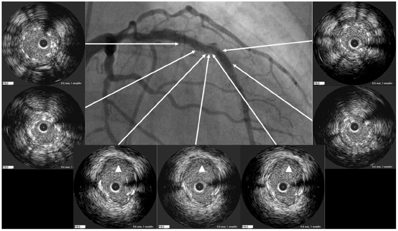
Fig. 2 IVUS at follow-up angiography confirmed a complete stent fracture with coronary artery aneurysm (arrow heads). IVUS: intravascular ultrasound.

Fig. 3 Cardiac MRI revealed myocardial thinning with transmural delayed hyperenhancement (arrow) on the anteroseptal area.
Reference
-
1. Brilakis ES, Maniu C, Wahl M, Barsness G. Unstable angina due to stent fracture. J Invasive Cardiol. 2004. 16:545.2. Sianos G, Hofma S, Ligthart JM, et al. Stent fracture and resenosis in the drug-eluting stent era. Catheter Cardiovasc Interv. 2004. 61:111–116.3. Min PK, Yoon YW, Kwon HM. Delayed strut fracture of sirolimus-eluting stent: a significant problem or an occasional observation? Int J Cardiol. 2006. 106:404–406.4. Surmely JF, Kinoshita Y, Dash D, et al. Stent strut fracture-induced restenosis in a bifurcation lesion treated with the crush stenting technique. Circ J. 2006. 70:936–938.5. Farb A, Burke AP, Kolodgie FD, Virmani R. Pathological mechanisms of fatal late coronary stent thrombosis in humans. Circulation. 2003. 108:1701–1706.6. Bae JH, Hyun DW, Kim KY, Yoon Hj, Nakamura S. Drug-eluting stent strut fracture as a cause of restenosis. Korean Circ J. 2005. 35:787–789.7. Kim JS, Yoon YW, Hong BK, et al. Delayed stent fracture after successful sirolimus-eluting stent (Cypher®) implantation. Korean Circ J. 2006. 36:443–449.8. Kim EJ, Rha SW, Wani SP, et al. Coronary stent fracture and restenosis in the drug-eluting stent era: do we have clues of management? Int J Cardiol. 2007. 120:417–419.9. Joner M, Finn AV, Farb A, et al. Pathology of drug-eluting stents in humans: delayed healing and late thrombotic risk. J Am Coll Cardiol. 2006. 48:193–202.
- Full Text Links
-
- Actions
-
Cited
- CITED
-
- Close
- Share
-
- Similar articles
-
- A Case of Coronary Artery Aneurysm after Sirolimus-Eluting Stent Implantation
- Complete Fracture of Sirolimus-Eluting Stent in a Saphenous Vein Graft to Left Anterior Descending Artery
- Drug-Eluting Stent Strut Fracture as a Cause of Restenosis
- Very Late Stent Thrombosis Related to Fracture of a Sirolimus-Eluting Stent
- A Case of Huge Coronary Aneurysm After Implantation of a Sirolimus-Eluting Stent
