Systolic Long Axis Function of the Left Ventricle, as Assessed by 2-D Strain, is Reduced in the Patients Who Have Diastolic Dysfunction and a Normal Ejection Fraction
- Affiliations
-
- 1Division of Cardiology, Cardiac and Vascular Center, Department of Medicine, Samsung Medical Center, Sungkyunkwan University School of Medicine, Seoul, Korea. parksmc@gmail.com
- KMID: 2225786
- DOI: http://doi.org/10.4070/kcj.2008.38.5.250
Abstract
-
BACKGROUND AND OBJECTIVES: Echocardiographic evaluation of the long axis left ventricle (LV) function has been reported to be useful for understanding heart failure in those patients with a preserved ejection fraction (EF). The global and segmental peak LV systolic longitudinal strain (PSLS), as determined by the 2D speckle tracking method, may be related with the conventional diastolic parameters. We sought to determine whether the PSLS could reveal LV systolic dysfunction in those patients who have a normal EF and diastolic dysfunction.
SUBJECTS AND METHODS
A total of 168 patients who underwent a routine echocardiographic examination were evaluated. Echocardiographic evaluations were performed and the patients were grouped according to the grade of their diastolic dysfunction. The global and segmental PSLS were analyzed off-line.
RESULTS
Measurements of the LV PSLS were successfully obtained in 83% of the patients. The mid and basal PSLS values were significantly lower in the patients with grade I and II diastolic dysfunction (-17.5+/-2.0% and -17.5+/-2.3%, respectively) versus the normal healthy controls (-20.6+/-1.9%, p<0.001). The mid and basal PSLS values were found to be well related to the early diastolic mitral annular velocity (r=0.510, p<0.001) and the left atrial volume index (r=-0.422, p<0.001).
CONCLUSION
The systolic LV long-axis function, as determined by 2D strain and especially in the mid and basal LV segments, is reduced in the patients with diastolic dysfunction in spite of their normal LV EF.
MeSH Terms
Figure
-
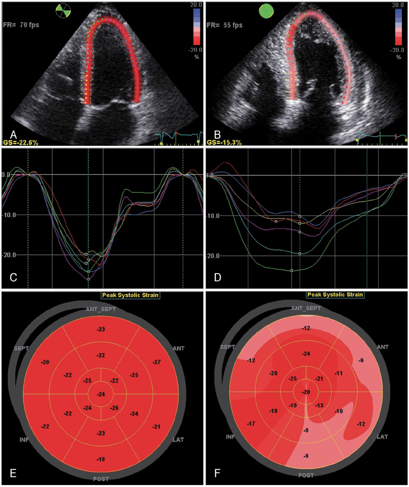
Fig. 1 Measurement of the peak systolic longitudinal strain (PSLS). The left column (A, C and E) represents a case from the normal group and the right column (B, D and F) represents a case from the diastolic dysfunction (DD) grade II group. The upper row (A and B) shows the speckle tracking process in the apical 4-chamber view. The middle row (C and D) shows the segmental strain curves. The lower row (E and F) shows the segmental PSLS in a bull's eye display. Note that in contrast to the normal patients, the PSLS in the DD grade II patients is reduced, and especially in the mid and basal segments. FR: frame rate, SEPT: septum, ANT_SEPT: anterior septum, INF: inferior wall, ANT: anterior wau, LAT: lateral wall, POST: posterior wall.
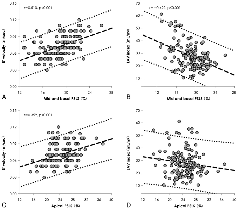
Fig. 2 Correlation plots between the peak systolic longitudinal strain (PSLS) and the diastolic parameters. As presented in A and B, the LAV index and E' were found to be well correlated with the mid and basal PSLS. Correlations between the apical PSLS and the LAV index or E' are presented in C and D. The PSLS values are presented as absolute values. E': peak early diastolic mitral annular velocity, LAV: left atrial volume.
Reference
-
1. Yip G, Wang M, Zhang Y, Fung JW, Ho PY, Sanderson JE. Left ventricular long axis function in diastolic heart failure is reduced in both diastole and systole: time for a redefinition? Heart. 2002. 87:121–125.2. Vinereanu D, Nicolaides E, Tweddel AC, Fraser AG. "Pure" diastolic dysfunction is associated with long-axis systolic dysfunction: implications for the diagnosis and classification of heart failure. Eur J Heart Fail. 2005. 7:820–828.3. Ha JW, Oh JK. The pathophysiology and diagnostic approaches for diastolic left ventricular dysfunction: a clinical perspective. Korean Circ J. 2005. 35:865–876.4. Park KH, Song JK, Suh IW, et al. The usefulness of 2-dimensional longitudinal strain for prediction of the postoperative left ventricular systolic function in patients with valvular heart disease causing volume overloading. Korean Circ J. 2006. 36:272–278.5. Reisner SA, Lysyansky P, Agmon Y, Mutlak D, Lessick J, Friedman Z. Global longitudinal strain: a novel index of left ventricular systolic function. J Am Soc Echocardiogr. 2004. 17:630–633.6. Jung HO, Youn HJ, Shin WS, et al. Differentiation of systolic and diastolic heart failure using strain and strain rate echocardiography. Korean Circ J. 2004. 34:1090–1098.7. Lowe BS, Tran H, Agler DA, Popovic ZB, Thomas JD, Grimm RA. Quantifying regional myocardial strain by online speckle tracking echocardiography: clinical feasibility and comparison with wall motion scoring. J Am Soc Echocardiogr. 2007. 20:584. abstract.8. Chinnaiyan KM, Alexander D, Maddens M, McCullough PA. Curriculum in cardiology: integrated diagnosis and management of diastolic heart failure. Am Heart J. 2007. 153:189–200.9. Solomon SD, Janardhanan R, Verma A, et al. Effect of angiotensin receptor blockade and antihypertensive drugs on diastolic function in patients with hypertension and diastolic dysfunction: a randomised trial. Lancet. 2007. 369:2079–2087.10. Lim TK, Ashrafian H, Dwivedi G, Collinson PO, Senior R. Increased left atrial volume index is an independent predictor of raised serum natriuretic peptide in patients with suspected heart failure but normal left ventricular ejection fraction: implication for diagnosis of diastolic heart failure. Eur J Heart Fail. 2006. 8:38–45.11. Lang RM, Bierig M, Devereux RB, et al. Recommendations for chamber quantification: a report from the American Society of Echocardiography's Guidelines and Standards Committee and the Chamber Quantification Writing Group, developed in conjunction with the European Association of Echocardiography, a branch of the European Society of Cardiology. J Am Soc Echocardiogr. 2005. 18:1440–1463.12. Pritchett AM, Jacobsen SJ, Mahoney DW, Rodeheffer RJ, Bailey KR, Redfield MM. Left atrial volume as an index of left atrial size: a population-based study. J Am Coll Cardiol. 2003. 41:1036–1043.13. Khouri SJ, Maly GT, Suh DD, Walsh TE. A practical approach to the echocardiographic evaluation of diastolic function. J Am Soc Echocardiogr. 2004. 17:290–297.14. Nishimura RA, Tajik AJ. Evaluation of diastolic filling of left ventricle in health and disease: Doppler echocardiography is the clinician's Rosetta Stone. J Am Coll Cardiol. 1997. 30:8–18.15. Pritchett AM, Mahoney DW, Jacobsen SJ, Rodeheffer RJ, Karon BL, Redfield MM. Diastolic dysfunction and left atrial volume: a population-based study. J Am Coll Cardiol. 2005. 45:87–92.16. Garcia MJ, Thomas JD, Klein AL. New Doppler echocardiographic applications for the study of diastolic function. J Am Coll Cardiol. 1998. 32:865–875.17. Vasan RS, Benjamin EJ, Levy D. Prevalence, clinical features and prognosis of diastolic heart failure: an epidemiologic perspective. J Am Coll Cardiol. 1995. 26:1565–1574.18. Petrie MC, Caruana L, Berry C, McMurray JJ. "Diastolic heart failure" or heart failure caused by subtle left ventricular systolic dysfunction? Heart. 2002. 87:29–31.19. Becker M, Bilke E, Kuhl H, et al. Analysis of myocardial deformation based on pixel tracking in two dimensional echocardiographic images enables quantitative assessment of regional left ventricular function. Heart. 2006. 92:1102–1108.20. Buckberg GD, Mahajan A, Jung B, Markl M, Hennig J, Ballester-Rodes M. MRI myocardial motion and fiber tracking: a confirmation of knowledge from different imaging modalities. Eur J Cardiothorac Surg. 2006. 29:Suppl 1. S165–S177.21. Kim HK, Sohn DW, Lee SE, et al. Assessment of left ventricular rotation and torsion with two-dimensional speckle tracking echocardiography. J Am Soc Echocardiogr. 2007. 20:45–53.22. Brucks S, Little WC, Chao T, et al. Contribution of left ventricular diastolic dysfunction to heart failure regardless of ejection fraction. Am J Cardiol. 2005. 95:603–606.23. Dokainish H, Zoghbi WA, Lakkis NM, et al. Incremental predictive power of B-type natriuretic peptide and tissue Doppler echocardiography in the prognosis of patients with congestive heart failure. J Am Coll Cardiol. 2005. 45:1223–1226.24. Svealv BG, Olofsson EL, Andersson B. Ventricular long axis function is of major importance for long-term survival in heart failure patients. Heart. 2008. 94:284–289.
- Full Text Links
-
- Actions
-
Cited
- CITED
-
- Close
- Share
-
- Similar articles
-
- Radionuclide Assessment of Cardiac Performance in Dilated Cardiomyopathy
- The Evaluation of Left and Right Ventricular Function by Radionuclide Ventriculography and Echocardiography in Dilated Cardiomyopathy
- Left Ventricular Diastolic Functions by M-Mode Echocardiogram in Essential Hypertensive Patients
- Differentiation of Systolic and Diastolic Heart Failure using Strain and Strain Rate Echocardiography
- Left Ventricular Diastolic Dysfunction: The What, When, and How
