Korean Circ J.
2008 Nov;38(11):596-600. 10.4070/kcj.2008.38.11.596.
Transient Left Ventricular Dysfunction After Percutaneous Patent Ductus Arteriosus Closure in Children
- Affiliations
-
- 1Department of Pediatrics, School of Medicine, Keimyung University, Daegu, Korea.
- 2Department of Pediatrics, College of Medicine, Pochon CHA University, Gumi, Korea.
- 3Department of Internal Medicine, School of Medicine, Kyungpook National University, Daegu, Korea.
- 4Department of Pediatrics, School of Medicine, Kyungpook National University, Daegu, Korea. mchyun@mail.knu.ac.kr
- KMID: 2225707
- DOI: http://doi.org/10.4070/kcj.2008.38.11.596
Abstract
- BACKGROUND AND OBJECTIVES
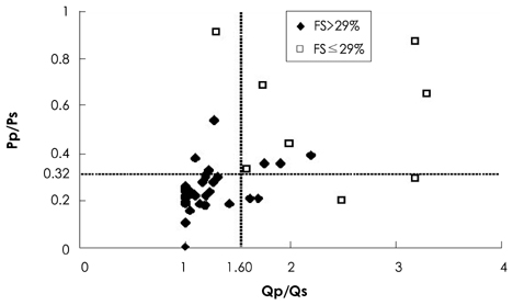
The goal of this study was to assess changes in left ventricular (LV) function and to identify pre-closure factors associated with LV dysfunction {fractional shortening (FS) below 29%} after transcatheter patent ductus arteriosus (PDA) closure. SUBJECTS AND METHODS: Forty-three pediatric patients with PDAs underwent cardiac catheterization for hemodynamic studies and intervention. Doppler echocardiography was performed at pre-closure, post-closure, and follow-up. RESULTS: S' and A' of the septum and mitral annulus were significantly decreased at post-closure and follow-up, respectively. In five of eight patients with Qp/Qs ratios over 1.60 and Pp/Ps ratios over 0.32 at pre-closure, the FS was decreased below 29% at post-closure. Qp/Qs ratio over 1.60 and Pp/Ps ratio over 0.32 at pre-closure had a sensitivity of 86% and a specificity of 84% for predicting FS to be below 29% at post-closure. CONCLUSION: Larger amounts of pre-closure left-to-right shunting and higher pulmonary artery pressure were associated with an increased likelihood of FS <29% after closure. The results of this study suggest that serial assessments of ventricular function are needed after PDA occlusion in patients with high Qp/Qs and Pp/Ps ratios.
MeSH Terms
Figure
Reference
-
1. Takahashi Y, Harada K, Ishida A, Tamura M, Tanaka T, Takada G. Changes in left ventricular volume and systolic function before and after the closure of ductus arteriosus in full-term infants. Early Hum Dev. 1996. 44:77–85.2. Galal MO, Amin M, Hussein A, Kouatli A, Al-Ata J, Jamjoom A. Left ventricular dysfunction after closure of large patent ductus arteriosus. Asian Cardiovasc Thorac Ann. 2005. 13:24–29.3. Masura J, Tittel P, Gavora P, Podnar T. Long-term outcome of transcatheter patent ductus arteriosus closure using Amplatzer duct occluders. Am Heart J. 2006. 151:755.4. Thanopoulos BD, Hakim FA, Hiari A, et al. Further experience with transcatheter closure of the patent ductus arteriosus using the Amplatzer duct occluder. J Am Coll Cardiol. 2000. 35:1016–1021.5. Faella HJ, Hijazi ZM. Closure of the patent ductus arteriosus with the amplatzer PDA device: immediate results of the international clinical trial. Catheter Cardiovasc Interv. 2000. 51:50–54.6. Bilkis AA, Alwi M, Hasri S, et al. The Amplatzer duct occluder: experience in 209 patients. J Am Coll Cardiol. 2001. 37:258–261.7. Hofbeck M, Bartolomaeus G, Buheitel G, et al. Safety and efficacy of interventional occlusion of patent ductus arteriosus with detachable coils: a multicentre experience. Eur J Pediatr. 2000. 159:331–337.8. Alwi M, Kang LM, Samion H, Latiff HA, Kandavel G, Zambahari R. Transcatheter occlusion of native persistent ductus arteriosus using conventional Gianturco coils. Am J Cardiol. 1997. 79:1430–1432.9. Her K, Lee JW, Shin HK, Won YS. Video-assisted thoracoscopic surgery for patent ductus arteriosus. Korean Circ J. 2004. 34:978–982.10. Galal MO, Arfi MA, Nicole S, Payot M, Hussain A, Qureshi S. Left ventricular systolic dysfunction after transcatheter closure of a large patent ductus arteriosus. J Coll Physicians Surg Pak. 2005. 15:723–725.11. Steinherz LJ, Graham T, Hurwitz R, et al. Guidelines for cardiac monitoring of children during and after anthracycline therapy. Pediatrics. 1992. 89:942–949.12. Porstmann W, Wierny L, Warnke H. The closure of the patent ductus arteriosus without thoracotomy (preliminary report). Thoraxchir Vask Chir. 1967. 15:199–203.13. Masura J, Walsh KP, Thanopoulous B, et al. Catheter closure of moderate- to large-sized patent ductus arteriosus using the new Amplatzer duct occluder: immediate and short-term results. J Am Coll Cardiol. 1998. 31:878–882.14. Park SS, Kim JH, Kwon MJ, Jung SW, Kim JK, Lee JH. Postnatal change of left ventricular contractility and volume in healthy term infants with ductus arteriosus. Korean Circ J. 2000. 30:1423–1429.15. Eerola A, Jokinen E, Boldt T, Pihkala J. The influence of percutaneous closure of patent ductus arteriosus on left ventricular size and function: a prospective study using two- and three-dimensional echocardiography and measurements of serum natriuretic peptides. J Am Coll Cardiol. 2006. 47:1060–1066.16. Jeong YH, Yun TJ, Song JM, et al. Left ventricular remodeling and change of systolic function after closure of patent ductus arteriosus in adults: device and surgical closure. Am Heart J. 2007. 154:436–440.17. Pauliks LB, Chan KC, Chang D, et al. Regional myocardial velocities and isovolumic contraction acceleration before and after device closure of atrial septal defects: a color tissue Doppler study. Am Heart J. 2005. 150:294–301.18. Dalsgaard M, Snyder EM, Kjaergaard J, Johnson BD, Hassager C, Oh JK. Isovolumic acceleration measured by tissue Doppler echocardiography is preload independent in healthy subjects. Echocardiography. 2007. 24:572–579.
- Full Text Links
- Actions
-
Cited
- CITED
-
- Close
- Share
- Similar articles
-
- Nonsurgical closure of patent ductus arteriosus with the rashkind PDA occluder system
- Percutaneous Forceps Retrieval of an Embolized Amplatzer Duct Occluder
- PDA Clipping by Using 2mm Thoracoscope
- Three Cases of Hemolysis After Transcatheter Closure of A Patent Ductus Arteriosus
- Occlusion of the Patent Ductus Arteriosus with Cook Detachable Coil


