Intravascular Ultrasound-Guided Troubleshooting in a Large Hematoma Treated With Fenestration Using a Cutting Balloon
- Affiliations
-
- 1Division of Cardiology, Department of Internal Medicine, Cardiac and Vascular Center, Samsung Medical Center, Sungkyunkwan University School of Medicine, Seoul, Korea. choijinh@skku.edu
- KMID: 2225694
- DOI: http://doi.org/10.4070/kcj.2009.39.4.171
Abstract
- Intramural hematoma formation is not a well-studied complication of percutaneous coronary intervention. We describe a patient with stable angina who developed an intramural hematoma during elective percutaneous coronary intervention (PCI) in the right coronary artery (RCA). Total occlusion with dense dye staining developed a long way from the distal RCA, near the posterior descending artery bifurcation site. The true lumen was compressed by the enlarged, tense, false lumen. The patient was successfully treating with intravascular ultrasound-guided fenestration using a cutting balloon, and a stent was implanted in the distal RCA.
Keyword
MeSH Terms
Figure
-
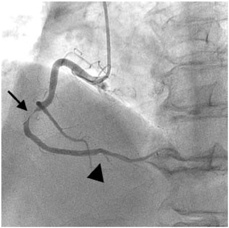
Fig. 1 Elective coronary angiography. The arrow indicates focal stenosis leading to 90% luminal narrowing in the mid-right coronary artery. The arrowhead indicates 99% luminal narrowing of the posterior descending artery ostium.

Fig. 2 Angiographic view of new narrowing that formed after stenting. Arrows indicate total occlusion and dye staining far from the distal right coronary artery, near the posterior descending artery bifurcation site.

Fig. 3 Site of the intramural hematoma that formed after stenting. Intravascular ultrasound revealed total compression of the distal right coronary artery lumen, which was caused by a huge hematoma (arrowheads).
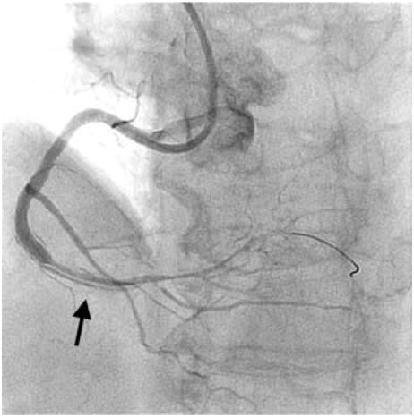
Fig. 4 Coronary angiogram after cutting balloon fenestration between the true lumen and the hematoma.
Reference
-
1. Maehara A, Mintz GS, Bui AB, et al. Incidence, morphology, angiographic findings, and outcomes of intramural hematomas after percutaneous coronary interventions. Circulation. 2002. 105:2037–2042.2. Hirose M, Kobayashi Y, Kreps EM, et al. Luminal narrowing due to intramural hematoma shift from left anterior descending coronary artery to left circumflex artery. Catheter Cardiovasc Interv. 2004. 62:461–465.3. Murphy DA, Craver JM, King SB 3rd. Distal coronary artery dissection following percutaneous transluminal coronary angioplasty. Ann Thorac Surg. 1984. 37:473–478.4. Zack PM, Ischinger T. Late progression of an asymptomatic intimal tear to occlusive coronary artery dissection following angioplasty. Cathet Cardiovasc Diagn. 1985. 11:41–48.5. Block PC, Myer RK, Stertzer S, Fallon JT. Morphology after transluminal angioplasty in human beings. N Engl J Med. 1981. 305:382–385.6. Stauffer JC, Sigwart U, Goy JJ, Kappenberger L. Milking dissection: an unusual complication of emergency coronary artery stenting for acute occlusion. Am Heart J. 1991. 121:1539–1542.7. Shirodaria C, van Gaal WJ, Banning AP. A bleeding kiss: intramural haematoma secondary to balloon angioplasty. Cardiovasc Ultrasound. 2007. 5:21.8. Song JK, Kang DH, Lee KM, et al. Two cases of aortic intramural hematoma diagnosed with transesophageal echocardiography. Korean Circ J. 1994. 24:904–909.9. van der Lugt A, Gussenhoven EJ, von Birgelen C, Tai JA, Pieterman H. Failure of intravascular ultrasound to predict dissection after balloon angioplasty by using plaque characteristics. Am Heart J. 1997. 134:1075–1081.10. Farb A, Virmani R, Atkinson JB, Kolodgie FD. Plaque morphology and pathologic changes in arteries from patients dying after coronary balloon angioplasty. J Am Coll Cardiol. 1990. 16:1421–1429.11. Lee RT, Kamm RD. Vascular mechanics for the cardiologist. J Am Coll Cardiol. 1994. 23:1289–1295.12. Waller BF. The eccentric coronary atherosclerotic plaque: morphologic observations and clinical relevance. Clin Cardiol. 1989. 12:14–20.13. Sheris SJ, Canos MR, Weissman NJ. Natural history of intravascular ultrasound-detected edge dissections from coronary stent deployment. Am Heart J. 2000. 139:59–63.14. Werner GS, Figulla HR, Grosse W, Kreuzer H. Extensive intramural hematoma as the cause of failed coronary angioplasty: diagnosis by intravascular ultrasound and treatment by stent implantation. Cathet Cardiovasc Diagn. 1995. 36:173–178.15. Werner GS, Diedrich J, Kreuzer H. Sonographic and angiographic features of intramural hematoma as a cause of failed coronary angioplasty. J Invasive Cardiol. 1996. 8:208–214.16. Mintz GS, Nissen SE, Anderson WD, et al. American College of Cardiology Clinical Expert Consensus Document on Standards for the Acquisition, Measurement, and Reporting of Intravascular Ultrasound Studies: a report of the American College of Cardiology Task Force on Clinical Expert Consensus Documents. J Am Coll Cardiol. 2001. 37:1478–1492.17. Mahr P, Ge J, Haude M, Gorge G, Erbel R. Extramural vessel wall hematoma causing a reduced vessel diameter after coronary stenting: diagnosis by intravascular ultrasound and treatment by stent implantation. Cathet Cardiovasc Diagn. 1998. 43:438–443.18. Fowlkes JB, Strieter RM, Downing LJ, et al. Ultrasound echogenicity in experimental venous thrombosis. Ultrasound Med Biol. 1998. 24:1175–1182.19. Sawada T, Shite J, Shinke T, et al. Persistent malapposition after implantation of sirolimus-eluting stent into intramural coronary hematoma: optical coherence tomography observations. Circ J. 2006. 70:1515–1519.20. Ahn SG, Tahk SJ, Choi JH, et al. Spontaneous coronary artery dissection manifested during ergonovine test and treated with intravascular ultrasound guided stenting: a case report. Korean Circ J. 2005. 35:264–268.
- Full Text Links
-
- Actions
-
Cited
- CITED
-
- Close
- Share
-
- Similar articles
-
- A Case Report of Percutaneous Fenestration of the Intimal Flap for Limb Ischemia in the Aortic Dissection
- Ultrasound-guided Evacuation of Spontaneous Intracerebral Hemorrhage in Basal Ganglia
- Effectiveness of the Hugging Balloon Technique in Coronary Angioplasty for a Heavy, Encircling, Calcified Coronary Lesion
- Cutting-edge progress of intravascular ultrasound in lower-extremity vascular interventions
- Endoscopic ultrasound-guided portal vein coiling: troubleshooting interventional endoscopic ultrasonography
