Korean Circ J.
2009 Nov;39(11):494-498. 10.4070/kcj.2009.39.11.494.
Isolated Left Ventricular Noncompaction Cardiomyopathy Accompanied by Severe Mitral Regurgitation
- Affiliations
-
- 1Division of Cardiology, Department of Internal Medicine, The Catholic University of Korea, St. Mary's Hospital, Seoul, Korea. younhj@catholic.ac.kr
- 2Department of Internal Medicine, Daerim Saint Mary's Hospital, Seoul, Korea.
- 3Department of Chest Surgery, The Catholic University of Korea, St. Mary's Hospital, Seoul, Korea.
- KMID: 2225653
- DOI: http://doi.org/10.4070/kcj.2009.39.11.494
Abstract
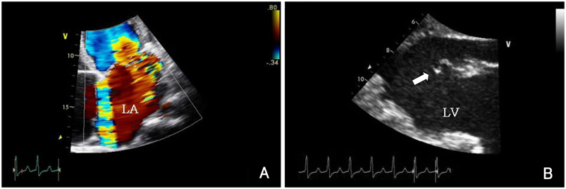
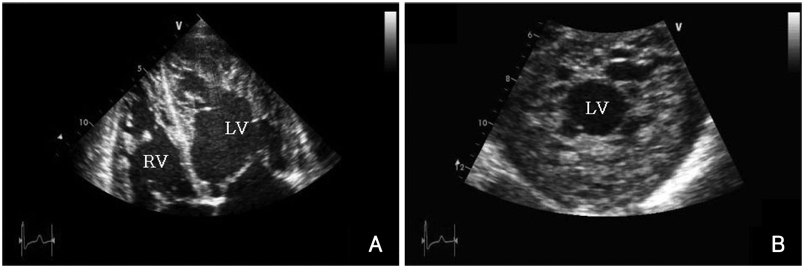
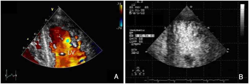
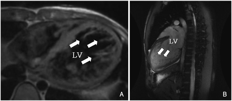
- Isolated left ventricular noncompaction cardiomyopathy (IVNC) is a cardiomyopathy thought to be caused by arrest of normal embryogenesis of the endocardium and myocardium. This abnormality is often associated with other congenital cardiac defects. A 21-year-old man presented to the emergency department with worsening exertional dyspnea during the previous 2 months. Two-dimensional and Doppler echocardiography revealed an enlarged left atrium (LA) and a markedly dilated left ventricle (LV) with preserved LV systolic function, severe mitral valve regurgitation, and prolapse due to chordae rupture. The myocardium of the LV and right ventricle (RV) had excessively prominent trabeculations and deep intertrabecular recesses. He is the first patient in Korea who has undergone mitral valve replacement surgery because of severe mitral valve regurgitation and prolapse due to chordae rupture accompanied by IVNC.
MeSH Terms
Figure
Reference
-
1. Chin TK, Perloff JK, Williams RG, Jue K, Mohrmann R. Isolated non-compaction of left ventricular myocardium: a study of eight cases. Circulation. 1990. 82:507–513.2. Richardson P, McKenna W, Bristow M, et al. Report of the 1995 World Health Organization/International Society and Federation of Cardiology Task Force on the definition and classification of cardiomyopathies. Circulation. 1996. 93:841–842.3. Agmon Y, Connolly HM, Olson LJ, Khadheria BK, Seward JB. Noncompaction of the ventricular myocardium. J Am Soc Echocardiogr. 1999. 12:859–863.4. Pignatelli R, McMahon JC, Dreyer WJ, et al. Clinical characterization of left ventricular noncompaction in children: a relatively common form of cardiomyopathy. Circulation. 2003. 108:2672–2678.5. Weiford BC, Subbarao VD, Mulhern KM. Noncompaction of the ventricular myocardium. Circulation. 2004. 109:2965–2971.6. Stamou SC, Lefrak EA, Athari FC, Burton NA, Massimiano PS. Heart transplantation in a patient with isolated noncompaction of the left ventricular myocardium. Ann Thorac Surg. 2004. 77:1806–1808.7. Conraads V, Paelinck B, Vorlat A, Goethals M, Jacobs W, Vrints C. Isolated non-compaction of the left ventricle: a rare indication for transplantation. J Heart Lung Transplant. 2001. 20:904–907.8. Engberding R, Bender F. Identification of a rare congenital anomaly of the myocardium by two-dimensional echocardiography: persistence of isolated myocardial sinusoids. Am J Cardiol. 1984. 53:1733–1734.9. Jenni R, Goebel N, Tartini R, Schneider J, Arbenz U, Oelz O. Persisting myocardial sinusoids of both ventricles as an isolated anomaly: echocardiographic, angiographic, and pathologic anatomical findings. Cardiovasc Intervent Radiol. 1986. 9:127–131.10. Jenni R, Oechslin EN, van der Loo B. Isolated ventricular noncompaction of the myocardium in adults. Heart. 2007. 93:11–15.11. Sasse-Klaassen S, Gerull B, Oechslin E, Jenni R, Thierfelder L. Isolated noncompaction of the left ventricular myocardium in the adult is an autosomal dominant disorder in the majority of patients. Am J Med Genet A. 2003. 119A:162–167.12. Lee SW, Koh MB, Hur WH, et al. A case of spongy myocardium initially manifested by ventricular tachycardia in adult. Korean J Med. 2003. 65:Suppl 3. S733–S737.13. Kim WS, Mang JH, Park SJ, et al. Two case of spongy myocardium detected in adult. J Korean Soc Echocardiogr. 2003. 11:108–113.14. Lim YJ, Kim JS, Kim IH, et al. A case of isolated noncompaction of the ventricular myocardium in an elderly patient. Korean J Med. 2007. 73:96–102.15. Ritter M, Oechslin E, Sütsch G, Attenhofer C, Schneider J, Jenni R. Isolated noncompaction of the myocardium in adults. Mayo Clin Proc. 1997. 72:26–31.16. Soler R, Rodriguez E, Monserrat L, Alvarez N. MRI of subendothelial perfusion deficits in isolated left ventricular noncompaction. J Comput Assist Tomogr. 2002. 26:373–375.17. Oechslin EN, Attenhofer-Jost C, Rojas JR, Kaufmann PA, Jenni R. Long-term follow-up of 34 adults with isolated left ventricular noncompaction: a distinct cardiomyopathy with poor prognosis. J Am Coll Cardiol. 2000. 36:493–500.
- Full Text Links
- Actions
-
Cited
- CITED
-
- Close
- Share
- Similar articles
-
- A Case of Left Ventricular Noncompaction Accompanying Fasciculo-Ventricular Accessory Pathway and Atrial Flutter
- Left Ventricular Noncompaction Associated with Hypertrophic Cardiomyopathy: Morphologic and Functional Evaluation with Multidetector CT
- Left ventricular function after mitral valve operation in congenital mitral regurgitation
- The Causing Factor of Mitral Regurgitation in Hypertrophic Cardiomyopathy
- Noncompaction of Ventricular Myocardium Involving the Right Ventricle

