Korean Circ J.
2009 Dec;39(12):532-537. 10.4070/kcj.2009.39.12.532.
Characteristics of Myocardial Deformation and Rotation in Subjects With Diastolic Dysfunction Without Diastolic Heart Failure
- Affiliations
-
- 1Department of Cardiology, Fatima General Hospital, Daegu, Korea. Augustjbc@yahoo.co.kr
- KMID: 2225641
- DOI: http://doi.org/10.4070/kcj.2009.39.12.532
Abstract
- BACKGROUND AND OBJECTIVES
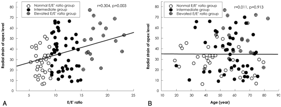
There have been very few pathophysiologic studies on isolated diastolic dysfunction. We hypothesized that the characteristics of isolated diastolic dysfunction would be located, on the clinical continuum, between those of a normal heart and diastolic heart failure. SUBJECTS AND METHODS: We enrolled 102 subjects who had no history of overt symptoms of heart failure and who had a left ventricular ejection fraction of more than 50%. They were examined for myocardial deformation and rotation using the two-dimensional speckle tracking image (2D-STI) technique. RESULTS: The circumferential strains and radial strain at the apical level (RS(apex)) were related to the ratio of the transmitral early peak velocity over the early diastolic mitral annulus velocity (E/E'). After adjustment for age, the RS(apex) showed a positive relationship with the E/E' ratio; whereas, the circumferential strains did not. Instead, the circumferential strains demonstrated a significant correlation with age. Basal rotation and left ventricular (LV) torsion were also related to age, but had no relationship with the E/E' ratio. However, as the E/E' ratio value increased, systolic mitral annular velocity decreased. CONCLUSION: Except for the RS(apex), LV myocardial deformation and rotation did not vary with the degree of E/E' ratio elevation when there was no associated diastolic heart failure. Additionally, in clinical situations such as isolated diastolic dysfunction, the advancement of age has a relatively greater influence on characteristics of LV myocardial deformation and rotation rather than on the E/E' ratio.
MeSH Terms
Figure
Reference
-
1. Kapila R, Mahajan RP. Diastolic dysfunction. CEACCP. 2009. 9:29–33.2. McKee PA, Castelli WP, McNamara PM, Kannel WB. The natural history of congestive heart failure: the Framingham study. N Engl J Med. 1971. 285:1441–1446.3. Quinones MA, Otto CM, Stoddard M, Waggoner A, Zoghbi WA. Recommendations for quantification of Doppler echocardiography: a report from the Doppler Quantification Task Force of the Nomenclature and Standards Committee of the American Society of Echocardiography. J Am Soc Echocardiogr. 2002. 15:167–184.4. Lee HJ, Kim BS, Kim JH, et al. Age-related changes in left ventricular torsion as assessed by 2-dimensional ultrasound speckle tracking imaging. Korean Circ J. 2008. 38:529–535.5. European Study Group on Diastolic Heart failure. How to diagnose diastolic heart failure. Eur Heart J. 1998. 19:990–1003.6. Vasan RS, Benjamin EJ, Levy D. Prevalence, clinical features and prognosis of diastolic heart failure. J Am Coll Cardiol. 2005. 26:1565–1574.7. Kass DA. Assessment of diastolic dysfunction: invasive modalities. Cardiol Clin. 2000. 18:571–586.8. Zile MR, Brutsaert DL. New concepts in diastolic dysfunction and diastolic heart failure: part I. diagnosis, prognosis, and measurements of diastolic function. Circulation. 2002. 105:1387–1393.9. Borlaug BA, Kass DA. Mechanisms of diastolic dysfunction in heart failure. Trends Cardiovasc Med. 2006. 16:273–279.10. Garcia MJ, Ares MA, Asher C, Rodriguez L, Vandervoort P, Thomas JD. An index of early left ventricular filling that combined with pulsed Doppler peak E velocity may estimate capillary wedge pressure. J Am Coll Cardiol. 1997. 29:448–454.11. Oki T, Tabata T, Yamata H, et al. Clinical application of pulsed tissue Doppler imaging for assessing abnormal left ventricular relaxation. Am J Cardiol. 1997. 79:921–928.12. Nishimura RA, Tajik AJ. Evaluation of diastolic filling of left ventricle in health and disease: doppler echocardiography is the clinician's Rosetta stone. J Am Coll Cardiol. 1997. 30:8–18.13. Nagueh SF, Middleton JK, Kopelen HA, Zoghbi WA, Quiñones MA. Doppler tissue imaging: a non-invasive technique for evaluation of left ventricular relaxation and estimation of filling pressures. J Am Coll Cardiol. 1997. 30:1527–1533.14. Ommen SR, Nishimura RA, Appleton CP, et al. Clinical utility of Doppler echocardiography and tissue Doppler imaging in the estimation of left ventricular filling pressures: a comparative simultaneous Doppler-catheterization study. Circulation. 2000. 102:1788–1794.15. Kim YJ, Sohn DW. Mitral annulus velocity in the estimation of left ventricular filling pressure: prospective study in 200 patients. J Am Soc Echocardiogr. 2000. 13:980–985.16. Yu CM, Lin H, Yang H, Kong SL, Zhang Q, Lee SW. Progression of systolic abnormalities in patients with "isolated" diastolic heart failure and diastolic dysfunction. Circulation. 2002. 105:1195–1201.17. Vinereanu D, Nicolaides E, Tweddel AC, Fraser AG. 'Pure' diastolic dysfunction is associated with long-axis systolic dysfunction: implications for the diagnosis and classification of heart failure. Eur J Heart Fail. 2005. 7:820–828.18. Wang J, Khoury DS, Yue Y, Torre-Amione G, Nagueh SF. Preserved left ventricular twist and circumferential deformation, but depressed longitudinal and radial deformation in patients with diastolic heart failure. Eur heart J. 2008. 29:1283–1289.19. Takeuchi M, Nakai H, Kokumai M, Nishikage T, Otani S, Lang RM. Age-related changes in left ventricular twist assessed by two-dimensional speckle-tracking imaging. J Am Soc Echocardiogr. 2006. 19:1077–1084.20. Kocica MJ, Corno AF, Carreras-Costa F, et al. The helical ventricular myocardial band: global, three-dimensional, functional architecture of the ventricular myocardium. Eur J Cardiothorac Surg. 2006. 29:Suppl 1. S21–S40.21. Matsuzaki M, Tanaka N, Toma Y, et al. Effect of changing afterload and inotropic states on inner and outer ventricular wall thickening. Am J Physiol. 1992. 263:H109–H116.22. Akagawa E, Murata K, Tanaka N, et al. Augmentation of left ventricular apical endocardial rotation with inotropic stimulation contributes to increased left ventricular torsion and radial strain in normal subjects: quantitative assessment utilizing a novel automated tissue tracking technique. Circ J. 2007. 71:661–668.
- Full Text Links
- Actions
-
Cited
- CITED
-
- Close
- Share
- Similar articles
-
- The Pathophysiology and Diagnostic Approaches for Diastolic Left Ventricular Dysfunction: A Clinical Perspective
- Therapeutic Strategies for Diastolic Dysfunction: A Clinical Perspective
- Perioperative management of left ventricular diastolic dysfunction and heart failure: an anesthesiologist's perspective
- Simple and Practical Way of Assessing Diastolic Function: Diastolic Heart Failure Revisited
- Left Ventricular Dyssynchrony in Patients Showing Diastolic Dysfunction without Overt Symptoms of Heart Failure


