Assessment of Epicardial Fat Volume With Threshold-Based 3-Dimensional Segmentation in CT: Comparison With the 2-Dimensional Short Axis-Based Method
- Affiliations
-
- 1Department of Radiology, Gyeongsang National University Hospital, College of Medicine, Gyeongsang National University, Jinju, Korea.
- 2Department of Radiology, Seoul St. Mary's Hospital, College of Medicine, The Catholic University of Korea, Seoul, Korea. cjy0122@yuhs.ac
- 3Department of Cardiology, Seoul St. Mary's Hospital, College of Medicine, The Catholic University of Korea, Seoul, Korea.
- KMID: 2225176
- DOI: http://doi.org/10.4070/kcj.2010.40.7.328
Abstract
- BACKGROUND AND OBJECTIVES
We aimed to assess the usefulness of a threshold-based, 3-dimensional (3D) segmentation in comparison with the traditional 2-dimensional (2D) short axis-based method for measurement of epicardial fat volume with 64-slice multidetector computed tomography (MDCT).
SUBJECTS AND METHODS
One hundred patients (52 males; mean age, 58.36+/-11.0 years) who underwent coronary CT angiography were enrolled in this study. The epicardial fat volume was measured using the two methods. The existing method was the 2D short axis-based method and the new method was the threshold-based 3D segmentation. Pearson's correlation was used to compare the two measurement methods. We also assessed the relationship between the epicardial fat volume and coronary artery disease (CAD).
RESULTS
There were a strong correlation between the epicardial fat volumes determined using the two methods (r=0.956, p<0.001). The mean overestimation of epicardial fat volume by the threshold-based 3D method was 59.89+/-12.00% compared to the 2D short-axis based method. Using the 3D method, the epicardial fat volume was significantly higher in the CAD group than in the controls (165.07+/-48.22 cm3 vs. 108.39+/-48.03 cm3, p<0.001).
CONCLUSION
Threshold-based 3D segmentation is another easy and useful tool for measuring the epicardial fat volume.
MeSH Terms
Figure
-

Fig. 1 Epicardial fat area measurement using 2-dimensional short axis-based method as the known method. A: on the short axis view, the region of interest (ROI; red line) is drawn around the pericardium in order to determine the epicardial fat area (green). B: the epicardial fat area is automatically measured by a histogram-based analysis (CT attenuation range between -200 HU and -30 HU). The epicardial fat area of this case is 735 m2. HU: Hounsfield unit.
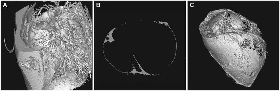
Fig. 2 Epicardial fat volume measurement using threshold-based 3-dimensional (3D) segmentation as the new method. A: the initial 3D volumetric data obtained not only intrathoracic fat, but also extrathoracic fat tissue (CT attenuation range between -200 HU and -30 HU). B: for the accurate measurement of the epicardial fat volume, we compared it to the corresponding 2-dimensional axial image. C: surface shaded display image demonstrates 3D epicardial fat volume (gray color). HU: Hounsfield unit.
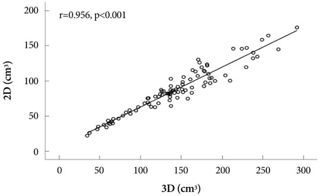
Fig. 3 Correlation dot plot of the epicardial fat volume in both methods. The correlation dot plot shows the strong correlation of each epicardial fat volumes measured using the two different methods (r=0.956, p<0.001); the x-axis indicates the 3D volume measurement (3D) and the y-axis denotes 2D short-axis based method (2D). 2D: 2-dimensional, 3D: 3-dimensional.
Reference
-
1. Taguchi R, Takasu J, Itani Y, et al. Pericardial fat accumulation in men as a risk factor for coronary artery disease. Atherosclerosis. 2001. 157:203–209.2. Djaberi R, Schuijf JD, van Werkhoven JM, Nucifora G, Jukema JW, Bax JJ. Relation of epicardial adipose tissue to coronary atherosclerosis. Am J Cardiol. 2008. 102:1602–1607.3. Gorter PM, de Vos AM, van der Graaf Y, et al. Relation of epicardial and pericoronary fat to coronary atherosclerosis and coronary artery calcium in patients undergoing coronary angiography. Am J Cardiol. 2008. 102:380–385.4. Jeong JW, Jeong MH, Yun KH, et al. Echocardiographic epicardial fat thickness and coronary artery disease. Circ J. 2007. 71:536–539.5. Ahn SG, Lim HS, Joe DY, et al. Relationship of epicardial adipose tissue by echocardiography to coronary artery disease. Heart. 2008. 94:e7.6. Rosito GA, Massaro JM, Hoffmann U, et al. Pericardial fat, visceral abdominal fat, cardiovascular disease risk factors, and vascular calcification in a community-based sample: the Framingham Heart Study. Circulation. 2008. 117:605–613.7. Galili O, Versari D, Sattler KJ, et al. Early experimental obesity is associated with coronary endothelial dysfunction and oxidative stress. Am J Physiol Heart Circ Physiol. 2007. 292:H904–H911.8. Fluchter S, Haghi D, Dinter D, et al. Volumetric assessment of epicardial adipose tissue with cardiovascular magnetic resonance imaging. Obesity. 2007. 15:870–878.9. Dey D, Suzuki Y, Suzuki S, et al. Automated quantitation of pericardiac fat from noncontrast CT. Invest Radiol. 2008. 43:145–153.10. Gorter PM, van Lindert AS, de Vos AM, et al. Quantification of epicardial and peri-coronary fat using cardiac computed tomography: reproducibility and relation with obesity and metabolic syndrome in patients suspected of coronary artery disease. Atherosclerosis. 2008. 197:896–903.11. Wheeler GL, Shi R, Beck SR, et al. Pericardial and visceral adipose tissues measured volumetrically with computed tomography are highly associated in type 2 diabetic families. Invest Radiol. 2005. 40:97–101.12. Sarin S, Wenger C, Marwaha A, et al. Clinical significance of epicardial fat measured using cardiac multislice computed tomography. Am J Cardiol. 2008. 102:767–771.13. Wang TD, Lee WJ, Shih FY, et al. Relations of epicardial adipose tissue measured by multidetector computed tomography to components of the metabolic syndrome are region-specific and independent of anthropometric indexes and intraabdominal visceral fat. J Clin Endocrinol Metab. 2009. 94:662–669.14. Kauczor HU, Heussel CP, Fischer B, Klamm R, Mildenberger P, Thelen M. Assessment of lung volumes using helical CT at inspiration and expiration: comparison with pulmonary function tests. AJR Am J Roentgenol. 1998. 171:1091–1095.15. Magnusson M, Lenz R, Danielsson PE. Evaluation of methods for shaded surface display of CT volumes. Comput Med Imaging Graph. 1991. 15:247–256.16. Juergens KU, Seifarth H, Range F, et al. Automated threshold-based 3D segmentation versus short-axis planimetry for assessment of global left ventricular function with dual-source MDCT. AJR Am J Roentgenol. 2008. 190:308–314.17. Cho H, Shin G, Lee J. Visceral fat accumulation in coronary artery disease. Korean Circ J. 1998. 28:740–748.18. Seo J, Kim DS, Kwon HM, et al. Severity of coronary artery disease and visceral fat obesity. Korean Circ J. 1998. 28:1176–1184.19. Tepe G, Wendel HP, Khorchidi S, et al. Thrombogenicity of various endovascular stent types: an in vitro evaluation. J Vasc Interv Radiol. 2002. 13:1029–1035.20. White CM, Sander S, Coleman CI, et al. Impact of epicardial anterior fat pad retention on postcardiothoracic surgery atrial fibrillation incidence: the AFIST-III Study. J Am Coll Cardiol. 2007. 49:298–303.
- Full Text Links
-
- Actions
-
Cited
- CITED
-
- Close
- Share
-
- Similar articles
-
- Semiautomatic Three-Dimensional Threshold-Based Cardiac Computed Tomography Ventricular Volumetry in Repaired Tetralogy of Fallot: Comparison with Cardiac Magnetic Resonance Imaging
- Hydrocephalus: Ventricular Volume Quantification Using Three-Dimensional Brain CT Data and Semiautomatic Three-Dimensional Threshold-Based Segmentation Approach
- Comparison between Three-Dimensional Navigator-Gated Whole-Heart MRI and Two-Dimensional Cine MRI in Quantifying Ventricular Volumes
- Usefulness of Two-dimensioanl CT & Three-dimensional CT in Blow-out Fracture
- Right Ventricular Mass Quantification Using Cardiac CT and a Semiautomatic Three-Dimensional Hybrid Segmentation Approach: A Pilot Study
