Korean Circ J.
2013 Apr;43(4):261-264. 10.4070/kcj.2013.43.4.261.
Three-Dimensional Angiography-Guided Percutaneous Transluminal Angioplasty for Distal Aorta and Bi-Iliac Chronic Total Occlusion
- Affiliations
-
- 1Cardiovascular Center, Korea University Guro Hospital, Seoul, Korea. swrha617@yahoo.co.kr
- KMID: 2224949
- DOI: http://doi.org/10.4070/kcj.2013.43.4.261
Abstract
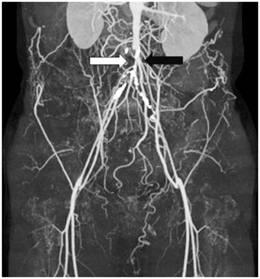
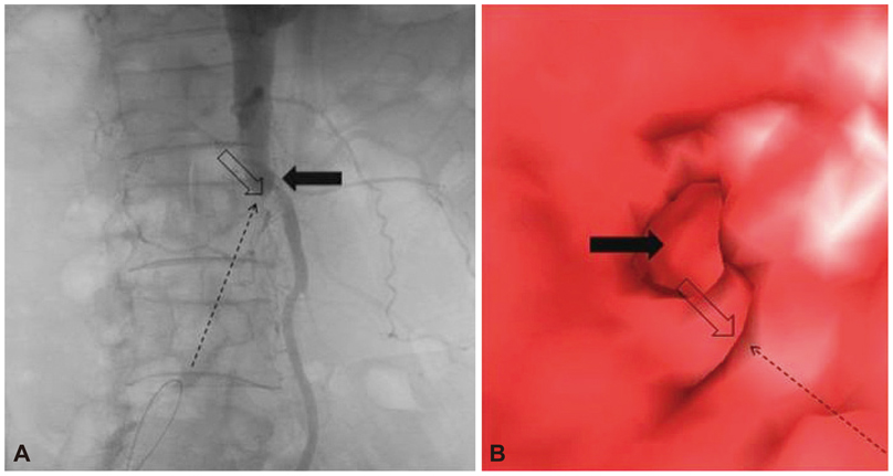
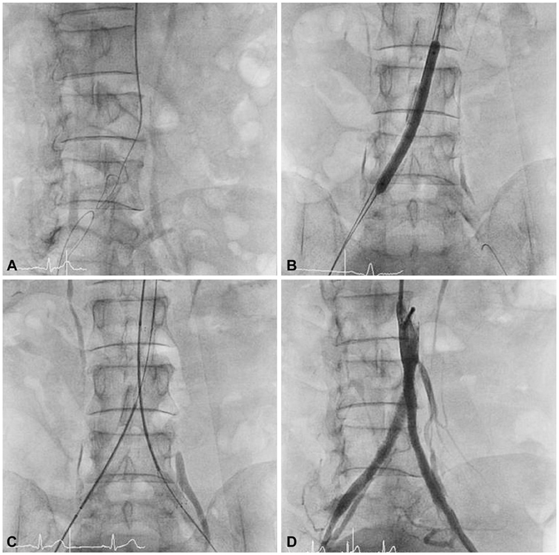
- Percutaneous recanalization of chronic total occlusions (CTOs) in peripheral arteries, especially TASC D classification including the distal aorta and both iliac arteries is still technically challenging. The conventional technique using standard guidewires and catheters guided by computed tomography and angiography can achieve a limited initial success, depending on lesion characteristics and operator's experience. A special imaging technique using 3-dimensional rotational angiography and spatio-temporal reconstruction with endoview for a better examination of the proximal stump, exact obstruction location, and distal stump direction in a stumpless lesion can be indispensable for successful intervention. We report a successful revascularization case of stumpless distal aorta and bi-iliac CTO guided by this specialized imaging technique.
Keyword
MeSH Terms
Figure
Reference
-
1. Gallagher KA, Meltzer AJ, Ravin RA, et al. Endovascular management as first therapy for chronic total occlusion of the lower extremity arteries: comparison of balloon angioplasty, stenting, and directional atherectomy. J Endovasc Ther. 2011. 18:624–637.2. van der Heijden FH, Eikelboom BC, Banga JD, Mali WP. Management of superficial femoral artery occlusive disease. Br J Surg. 1993. 80:959–963.3. Al-Ameri H, Shin V, Mayeda GS, et al. Peripheral chronic total occlusions treated with subintimal angioplasty and a true lumen re-entry device. J Invasive Cardiol. 2009. 21:468–472.4. Lee JB, Chang SG, Kim SY, et al. Assessment of three dimensional quantitative coronary analysis by using rotational angiography for measurement of vessel length and diameter. Int J Cardiovasc Imaging. 2012. 28:1627–1634.5. Kawasaki D, Tsujino T, Fujii K, Masutani M, Ohyanagi M, Masuyama T. Novel use of ultrasound guidance for recanalization of iliac, femoral, and popliteal arteries. Catheter Cardiovasc Interv. 2008. 71:727–733.6. Kawarada O, Yokoi Y, Takemoto K. Practical use of duplex echo-guided recanalization of chronic total occlusion in the iliac artery. J Vasc Surg. 2010. 52:475–478.7. Dvir D, Assali A, Kornowski R. Percutaneous coronary intervention for chronic total occlusion: novel 3-dimensional imaging and quantitative analysis. Catheter Cardiovasc Interv. 2008. 71:784–789.8. Pannu N, Wiebe N, Tonelli M. Alberta Kidney Disease Network. Prophylaxis strategies for contrast-induced nephropathy. JAMA. 2006. 295:2765–2779.
- Full Text Links
- Actions
-
Cited
- CITED
-
- Close
- Share
- Similar articles
-
- Endovascular Management of Acute Total Occlusion of the Aorta
- A case of percutaneous transluminal angioplasty in Takayasu arteritis
- Percutaneous Transluminal Angioplasty of a Stenosis of an Internal Mammary Artery Graft
- Impotence due to External Iliac Steal Syndrome: Treatment with Percutaneous Transluminal Angioplasty and Stent Placement
- Percutaneous Transluminal Angioplasty of Contralateral Iliac and Superficial Femoral Arteries via Graft Vessel in a Patient with FemoroFemoral Bypass Graft


