Anomalous Separate Origin of Left Anterior Descending Coronary Artery: Presented as Acute Anterior Myocardial Infarction
- Affiliations
-
- 1Department of Internal Medicine, Ulsan University College of Medicine, Gangneung Asan Hospital, Gangneung, Korea. drshin@ulsan.ac.kr
- KMID: 2224902
- DOI: http://doi.org/10.4070/kcj.2013.43.6.408
Abstract
- Coronary artery anomalies are rare presentations in primary percutaneous coronary interventions of acute myocardial infarction. Herein, we report the case of a 59-year-old man with acute anterior myocardial infarction who had anomalous separate origin of left anterior descending artery (LAD) and left circumflex artery (LCX) from the left coronary aortic sinus. Coronary angiography showed a normal right coronary artery and LCX, but no visualization of the LAD. After several unsuccessful attempts to cannulate the LAD, we found the LAD ostium located by the side of the LCX ostium. There was total occlusion at proxymal LAD. Coronary computed tomography angiography demonstrated the precise, separate origin of LAD and LCX from the left coronary aortic sinus.
MeSH Terms
Figure
-
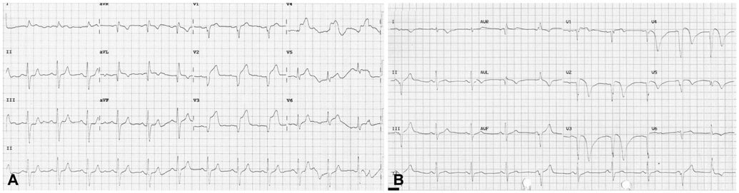
Fig. 1 Initial and follow-up electrocardiograms. A: the initial electrocardiogram showing ST-segment elevation on lead V 1-4. B: the electrocardiogram after primary percutaneous coronary intervention showing pathologic Q-wave and T-wave inversion on lead V 1-5.

Fig. 2 Coronary angiography and primary percutaneus coronary intervention. A: AP cranial projectoon 30 degree angle showing that the guidewire would not advance into the LAD ostium. B: LAO caudal protection marking the site of LAD stump (arrow). C: AP caudal view showing the separate LAD ostium directly originating from the left coronary aortic sinus. D: another AP caudal view showing residual 60% stenosis at proximal LAD after balloon angioplasty. AP: anterioposterior, LAD: left anterior descending artery, LAO: left anterior oblique, LCX: left circumflex artery.
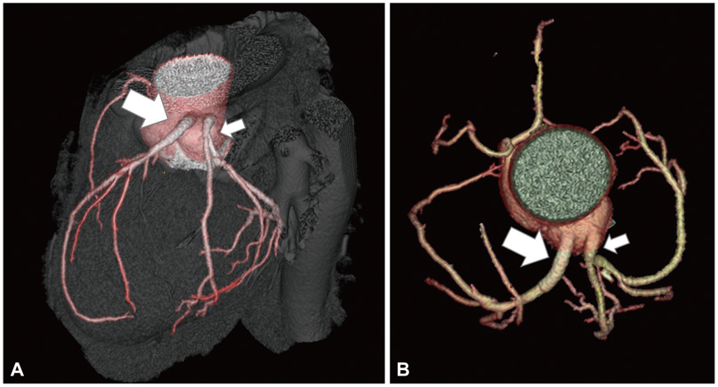
Fig. 3 Coronary CT angiography showing anomalous separate origin of LAD (large arrow) and LCX (small arrow) from the left coronary aortic sinus. A: LAO cranial view of a three-demensional volume rendered image. B: top view of a volume-rendered coronary tree image. LAD: left anterior descending artery, LAO: left anterior oblique, LCX: left circumflex artery.
Reference
-
1. Papadopoulos DP, Dalianis N, Benos I, Votteas V, Anagnostopoulou S. Anomalous origin of circumflex artery from right sinus of Valsalva: a rare cause of non-ST elevation syndrome. Int J Cardiol. 2007; 114:e105–e106.2. Kim JH, Ha GJ, Seong MJ, et al. Anomalous origin of the left circumflex coronary artery from the first diagonal branch presented as acute myocardial infarction. Korean Circ J. 2011; 41:612–614.3. Rohit M, Bagga S, Talwar KK. Double right coronary artery with acute inferior wall myocardial infarction. J Invasive Cardiol. 2008; 20:E37–E40.4. Cademartiri F, La Grutta L, Malagò R, et al. Prevalence of anatomical variants and coronary anomalies in 543 consecutive patients studied with 64-slice CT coronary angiography. Eur Radiol. 2008; 18:781–791.5. Taylor AJ, Rogan KM, Virmani R. Sudden cardiac death associated with isolated congenital coronary artery anomalies. J Am Coll Cardiol. 1992; 20:640–647.6. Serota H, Barth CW 3rd, Seuc CA, Vandormael M, Aguirre F, Kern MJ. Rapid identification of the course of anomalous coronary arteries in adults: the "dot and eye" method. Am J Cardiol. 1990; 65:891–898.7. Malagò R, D'Onofrio M, Brunelli S, et al. Anatomical variants and anomalies of the coronary tree studied with MDCT coronary angiography. Radiol Med. 2010; 115:679–692.
- Full Text Links
-
- Actions
-
Cited
- CITED
-
- Close
- Share
-
- Similar articles
-
- Acute Myocardial Infarction in a Patient with an Anomalous Origin of the Right Coronary Artery from the Left Anterior Descending Coronary Artery
- Anomalous Origin of the Left Coronary Artery Leading to Myocardial Infarction in a 14-year-old Boy
- Anomalous Origin of the Left Coronary Artery from the Right Sinus of Valsalva, which Presented as Acute Myocardial Infarction
- Anomalous Origin of the Right Coronary Artery from the Left Anterior Descending Artery: An Extremely Rare Variety of Single Coronary Artery
- Coronary-Pulmonary Fistulas Involving All Three Major Coronary Arteries Co-Existing With Myocardial Infarction
