Korean Circ J.
2013 Jun;43(6):384-390. 10.4070/kcj.2013.43.6.384.
Assesment of Myocardial Ischemia by Combination of Tissue Synchronisation Imaging and Dobutamine Stress Echocardiography
- Affiliations
-
- 1Department of Cardiology, Ataturk University, Erzurum, Turkey. mhakantas@gmail.com
- KMID: 2224899
- DOI: http://doi.org/10.4070/kcj.2013.43.6.384
Abstract
- BACKGROUND AND OBJECTIVES
Dobutamine stress echocardiography (DSE) is an important non-invasive imaging method for evaluating ischemia. However, wall motion interpretation can be impaired by the experience level of the interpreter and the subjectivity of the visual assessment. In our study we aimed to combine DSE and tissue syncronisation imaging to increase sensitivity for detecting ischemia.
SUBJECTS AND METHODS
50 patients with indications for DSE were included in the study. In 25 patients we found DSE positive for ischemia and in the other 25 patients we found it to be negative. The negative group was accepted as the control group. There was no significant difference in terms of risk factors and echocardiographic parameters between the two groups, except for wall motion scores. In both groups, left ventricular dyssychrony was accepted as the difference between time to peak systolic velocity (Ts) in the reciprocal four couple of non-apical segments at rest and during peak stress. Timings were corrected for heart rate. We compared the differences of the dyssynchronisation value at rest and during peak stress to determine the distinctions within the groups and between the groups of DSE positive and negative patients.
RESULTS
We found that stress and ischemia did not create any significant difference over the left intraventricular dyssynchrony with DSE, although at the segmenter level it prolonged the time to peak systolic velocity (p<0.05). These alterations did not show any significant difference between positive and negative DSE groups.
CONCLUSION
As a result, this segmenter dyssynchrony and the time to peak systolic velocity, which is corrected for heart rate, did not enhance any new value over DSE for detecting ischemia.
MeSH Terms
Figure
-
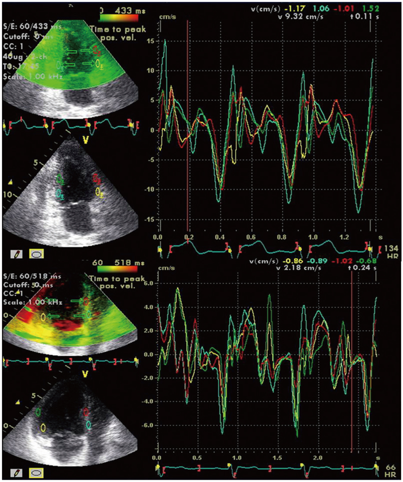
Fig. 1 Tissue synchronisation imaging and measurements of intraventricular dyssynchrony at 2 chamber and 4 chamber images.
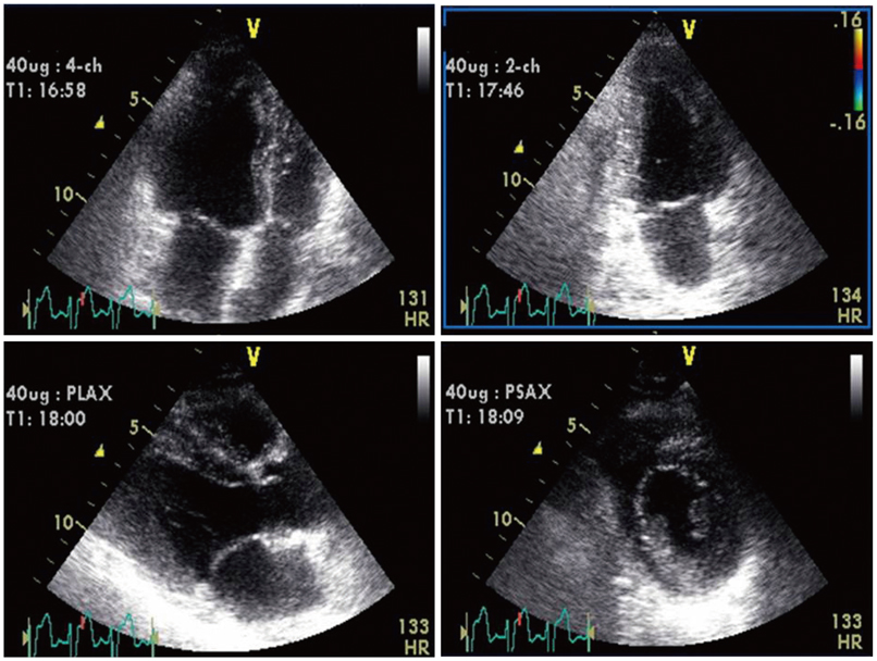
Fig. 2 Dobutamin stress echocardiography images at 40 mcgr/kg/minute.
Reference
-
1. Marwick TH. Stress echocardiography. Heart. 2003; 89:113–118.2. Gottdiener JS. Overview of stress echocardiography: uses, advantages, and limitations. Curr Probl Cardiol. 2003; 28:485–516.3. Picano E. Stress Echocardiography. 4th ed. Heidelberg, Germany: Springer-Verlag;2003.4. Corday E, Hajduczki I, O'Byrne GT, Kar S, Areeda J, Corday SR. Echocardiographic criteria to distinguish reversible from irreversible myocardial ischaemia. Eur Heart J. 1988; 9:Suppl F. 29–43.5. Tian JW, Du GQ, Ren M, Sun LT, Leng XP, Su YX. Tissue synchronization imaging of myocardial dyssynchronicity of the left ventricle in patients with coronary artery disease. J Ultrasound Med. 2007; 26:893–897.6. Kyriakides ZS, Manolis AG, Kolettis TM. The effects of ventricular asynchrony on myocardial perfusion. Int J Cardiol. 2007; 119:3–9.7. Schiller NB, Shah PM, Crawford M, et al. Recommendations for quantitation of the left ventricle by two-dimensional echocardiography. American Society of Echocardiography Committee on Standards, Subcommittee on Quantitation of Two-Dimensional Echocardiograms. J Am Soc Echocardiogr. 1989; 2:358–367.8. Dohi K, Pinsky MR, Suffoletto MS, Severyn DA, Gorcsan J III. A new rapid and simple index of mechanical dyssynchrony by colour-coded strain dyssynchrony imaging. J Am Coll Cardiol. 2005; 45:A289.9. Sade LE, Gorcsan J 3rd, Severyn DA, Edelman K, Katz WE. Usefulness of angle corrected tissue Doppler to assess segmental left ventricular function during dobutamine stress echocardiography in patients with and without coronary artery disease. Am J Cardiol. 2005; 96:141–147.10. Gorcsan J 3rd, Kanzaki H, Bazaz R, Dohi K, Schwartzman D. Usefulness of echocardiographic tissue synchronization imaging to predict acute response to cardiac resynchronization therapy. Am J Cardiol. 2004; 93:1178–1181.11. Leier CV, Unverferth DV. Drugs five years later. Dobutamine. Ann Intern Med. 1983; 99:490–496.12. Lafitte S, Bordachar P, Lafitte M, et al. Dynamic ventricular dyssynchrony: an exercise-echocardiography study. J Am Coll Cardiol. 2006; 47:2253–2259.13. Valzania C, Gadler F, Eriksson MJ, Olsson A, Boriani G, Braunschweig F. Electromechanical effects of cardiac resynchronization therapy during rest and stress in patients with heart failure. Eur J Heart Fail. 2007; 9:644–650.14. Kang SJ, Lim HS, Choi BJ, et al. The impact of exercise-induced changes in intraventricular dyssynchrony on functional improvement in patients with nonischemic cardiomyopathy. J Am Soc Echocardiogr. 2008; 21:948–953.15. De Sutter J, Van de Veire NR, Muyldermans L, et al. Prevalence of mechanical dyssynchrony in patients with heart failure and preserved left ventricular function (a report from the Belgian Multicenter Registry on dyssynchrony). Am J Cardiol. 2005; 96:1543–1548.16. Hummel JP, Lindner JR, Belcik JT, et al. Extent of myocardial viability predicts response to biventricular pacing in ischemic cardiomyopathy. Heart Rhythm. 2005; 2:1211–1217.17. Da Costa A, Thévenin J, Roche F, et al. Prospective validation of stress echocardiography as an identifier of cardiac resynchronization therapy responders. Heart Rhythm. 2006; 3:406–413.
- Full Text Links
-
- Actions
-
Cited
- CITED
-
- Close
- Share
-
- Similar articles
-
- Feasibility Study of Dobutamine Stress Transesophageal Echocardiography
- Comparison Study of Dipyridamole and Dobutamine Stress Echocardiography in Same Patients
- A Case of Giant Coronary Arteriovenous Fistula with Coronary Steal Demonstrated by Dobutamine Stress Echocardiography and (99m)Tc-MIBI SPECT
- Usefulness of Low Dose Dobutamine Stress Echocardiography in Patients with Acute Myocardial Infarction
- The Usefulness of Dobutamine Stress Echocardiography for Evaluation of Viable Myocardium in Hibernating Myocardium
